Science | Peking University Yulong Li lab develops the far-red dopamine sensor HaloDA1.0, enabling multiplex imaging of neurochemical networks in vivo
AAV9-hSyn-HaloDA1.0 Custom AAV | Peking University Yulong Li Lab Develops Far-Red Dopamine Sensor for Multiplex Imaging of Neurochemical Networks (Science)
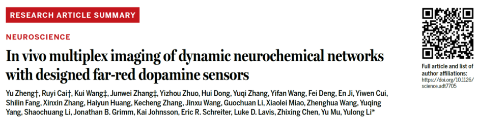
Paper title: In vivo multiplex imaging of dynamic neurochemical networks with designed far-red dopamine sensors
Journal: Science
Client: Professor Yulong Li's team, Peking University
Neuromodulators such as dopamine (DA) regulate brain function through interactions with acetylcholine (ACh), serotonin (5-HT) and others, but existing fluorescent sensors are limited to the green/red spectral range, making multicolor simultaneous imaging difficult. On June 5, 2025, the Yulong Li lab at Peking University, together with multiple domestic and international teams, published online in Science the paper entitled In vivo multiplex imaging of dynamic neurochemical networks with designed far-red dopamine sensors. The study developed a far-red dopamine sensor, HaloDA1.0, which combines cpHaloTag-based chemical dyes with the GRAB strategy, featuring high sensitivity, subsecond response kinetics, and a far-red to near-infrared spectral range. HaloDA1.0 can be used in combination with existing green and red fluorescent sensors to achieve multicolor imaging of dopamine together with other neuromodulators as well as calcium and cAMP in cultured neurons, brain slices and behaving animals, revealing the dynamic interaction mechanisms of dopamine within neurochemical networks and providing a new tool for dissecting complex brain functions.
Research Results
1. Development and in vitro characterization of the HaloDA1.0 sensor
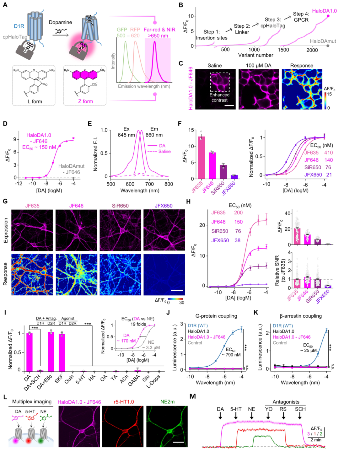
The research team replaced the third intracellular loop (ICL3) of the human D1 receptor (D1R) with an optimized circularly permuted HaloTag protein (cpHaloTag) combined with far-red dyes (mainly JF646). By screening more than 2,000 variants and optimizing the insertion site, linker sequences and key amino acid residues, they successfully developed the far-red dopamine sensor GRABHaloDA1.0 (abbreviated as HaloDA1.0). In vitro validation showed that HaloDA1.0 localized to the plasma membrane in HEK293T cells, and the fluorescence intensity rose transiently upon DA application, with a maximum fluorescence change (ΔF/F0) of about 900% and a half-maximal effective concentration (EC50) of 150 nM. By testing multiple rhodamine derivatives, the team found that different dyes could tune the sensor's spectrum, response amplitude and affinity. The team then characterized the pharmacological properties, kinetics, and coupling to downstream pathways of HaloDA1.0 expressed in HEK293T cells and cultured neurons, showing that the sensor has high sensitivity and specificity for DA, fast response kinetics, and no coupling to downstream signaling pathways. Importantly, when HaloDA1.0 (far-red), the red 5-HT sensor (r5-HT1.0) and the green NE sensor (NE2m) were co-expressed in cultured neurons, real-time simultaneous monitoring of three monoamine neurotransmitters was achieved with no significant crosstalk between the signals.
Development and characterization of the far-red dopamine sensor
2. HaloDA1.0 enables multiplex imaging in acute brain slices and zebrafish
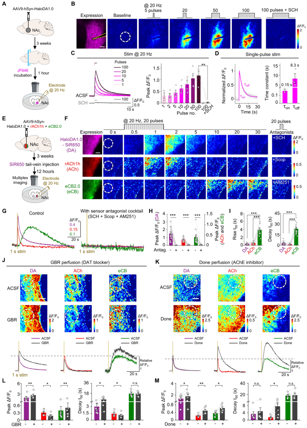
To evaluate whether HaloDA1.0 can detect endogenous DA release, an AAV expressing HaloDA1.0 was injected into the mouse nucleus accumbens (NAc), and acute brain slices were prepared 3 weeks later. After labeling with the JF646 dye, DA release was evoked by electrical stimulation. The results showed that 20 Hz electrical stimulation triggered a rapid increase in HaloDA1.0 fluorescence intensity, the response amplitude was positively correlated with the number of stimulation pulses, and DA release could be detected by even a single pulse; the D1R antagonist SCH completely blocked the fluorescence signal, confirming that the signal originated from endogenous DA release. Co-injection into the NAc of AAVs expressing HaloDA1.0, rACh1h and eCB2.0 enabled sensitive reporting of the endogenous dynamics of dopamine, acetylcholine and endocannabinoids evoked by electrical stimulation, revealing the distinct release kinetics of the three. In addition, the research team generated transgenic zebrafish co-expressing a green ATP probe, a red calcium probe and the far-red HaloDA1.0 probe. Combining three-color imaging, the researchers observed in real time, during electric shock stimulation and seizure-like behavior in zebrafish, synchronized release or activity of dopamine, extracellular ATP and neuronal calcium signals, with the three showing different decay rates during the signal dissipation phase.
Multiplex imaging with HaloDA1.0 in acute brain slices
3. HaloDA1.0 can detect the dynamic changes of multiple neuromodulators in living mice
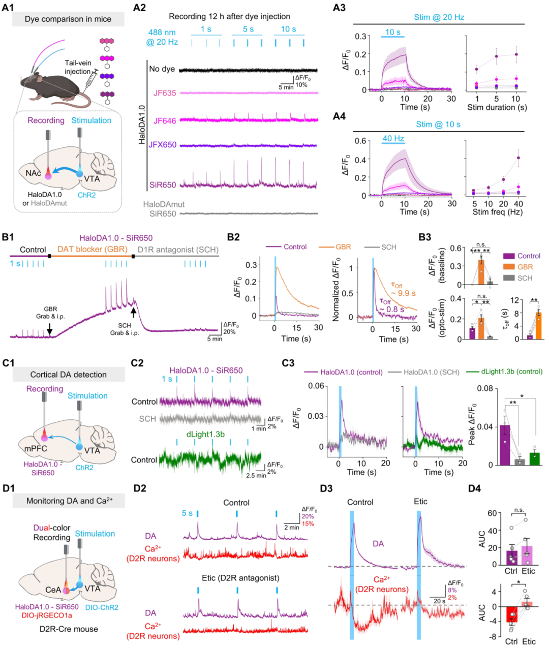
Using cpHaloTag-based sensors in mice requires delivering the dye to the mouse brain. To optimize the performance of HaloDA1.0, the research team systematically compared various far-red dyes in vivo. Rhodopsin-2 was expressed in the ventral tegmental area (VTA), and HaloDA1.0 was expressed in the NAc, which receives dense dopaminergic projections from the VTA. An AAV expressing the optogenetic activation protein ChR2 was injected into the mouse VTA, while HaloDA1.0 was expressed in the NAc or mPFC; different far-red dyes were injected via the tail vein to label the sensor, and DA dynamics were monitored by fiber photometry. The results showed that the SiR650-labeled HaloDA1.0 probe could specifically detect dopamine release induced by optogenetic activation of dopaminergic neurons both in the nucleus accumbens, which is rich in dopaminergic projections, and in the cerebral cortex, which is sparsely innervated. Furthermore, the research team used three fibers to simultaneously record the dynamics of DA, ACh and cAMP in D1 medium spiny neurons (D1-MSNs) of mice during spontaneous locomotion, sucrose reward, foot shock and cocaine injection. The results showed that during physiological behaviors, dopamine and acetylcholine jointly regulated cAMP through synergistic action, whereas under cocaine stimulation, their regulation of cAMP showed an antagonistic effect.
HaloDA1.0 can be used to detect endogenous DA release in freely moving mice
Conclusion
This study successfully developed the far-red dopamine probe HaloDA1.0, a novel sensor that sensitively and specifically reports the dynamic changes of dopamine across multiple systems, including cells, brain slices, zebrafish and mice. Used in combination with green and red fluorescent probes, HaloDA1.0 enables researchers to capture the changes of multiple neurochemical molecules simultaneously, providing an in-depth analysis of their spatiotemporal dynamics and regulatory relationships. This achievement is an important product of interdisciplinary collaboration; it both expands the boundaries of in vivo chemistry research and provides strong support for dissecting the brain's complex neurotransmitter regulatory networks.