Nat Commun | Wang Jing / Wu Zhaofa / Li Yulong teams develop a new HypnoS probe revealing intracellular adenosine dynamics and regulation
AAV-HypnoS Custom | Peking University Wang Jing and Li Yulong teams develop a new HypnoS probe revealing intracellular adenosine dynamics and regulation (Nature Communications, IF 14.7)
Paper title: A high-performance fluorescent sensor spatiotemporally reveals cell-type specific regulation of intracellular adenosine in vivo
Journal: Nature Communications (IF 14.7)
Collaborating partner: School of Pharmaceutical Sciences, Peking University (Wang Jing), in collaboration with the Institute of Genetics and Developmental Biology, Chinese Academy of Sciences (Wu Zhaofa) and the School of Life Sciences, Peking University (Li Yulong team)
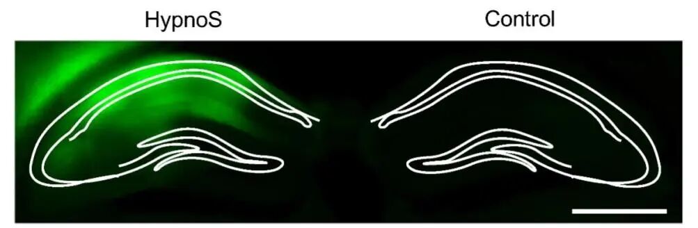
AAV2/9-hSyn-HypnoS mediates HypnoS expression in the hippocampus
Research Background
Adenosine (Ado) is a nucleoside that links intracellular metabolism with intercellular communication and plays an important role in processes such as the regulation of sleep and epilepsy. Although the function of extracellular Ado (eAdo) has been well established, the distribution and regulatory function of intracellular Ado (iAdo) remain poorly understood because of the lack of methods for detecting Ado in vivo. Recently, Professor Wang Jing of the School of Pharmaceutical Sciences, Peking University, together with Researcher Wu Zhaofa of the Institute of Genetics and Developmental Biology, Chinese Academy of Sciences, and Professor Li Yulong's team of the School of Life Sciences, Peking University, published the paper A high-performance fluorescent sensor spatiotemporally reveals cell-type specific regulation of intracellular adenosine in vivo in Nature Communications (IF 14.7). The study successfully developed HypnoS, the first genetically encoded fluorescent sensor for detecting intracellular adenosine, and, using a range of advanced in vivo imaging approaches, explored in depth the cell-type-specific spatiotemporal regulation of intracellular adenosine in living animals, unravelling the complex molecular regulation of adenosine homeostasis.
Research Results
1. HypnoS monitors iAdo dynamics
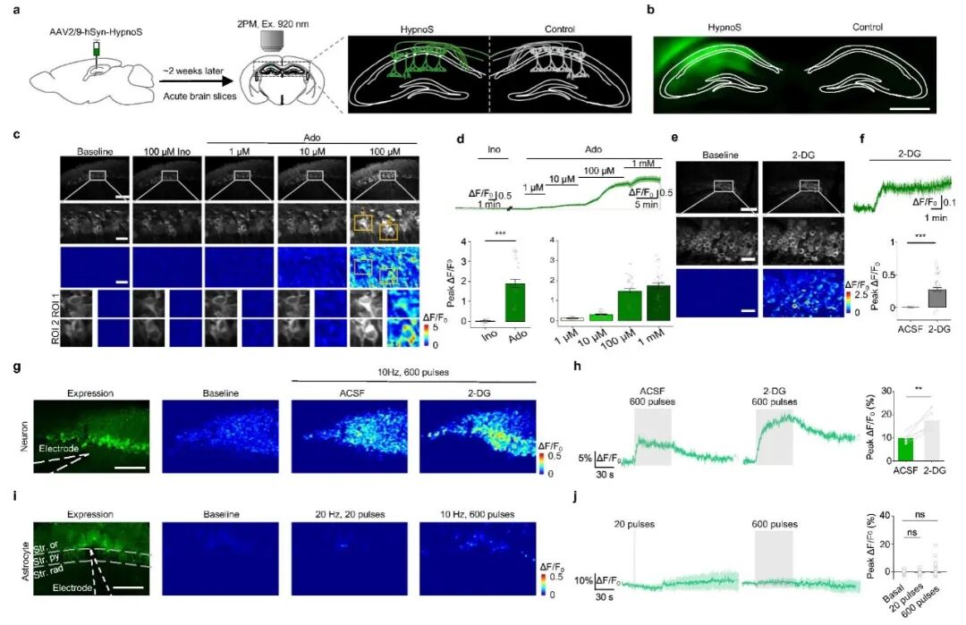
Based on a circularly permuted green fluorescent protein (cpEGFP) and an adenosine deaminase (PvADA), the authors developed the fluorescent sensor HypnoS for detecting intracellular adenosine and the control sensor HypnoS-mut, and demonstrated in mammalian cells that HypnoS is a sensitive and specific sensor for iAdo. To assess the sensitivity of HypnoS for monitoring iAdo dynamics in brain tissue, the authors used AAV2/9-hSyn-HypnoS (provided by WZBIO) to express HypnoS in mice, and found that HypnoS offers single-cell resolution, with iAdo levels observed to rise progressively in neurons in acute brain slices under 2-DG-induced metabolic suppression and electrical stimulation. In contrast, astrocytes showed smaller changes in iAdo levels in response to electrical stimulation, but responded to the application of 100 μM Ado. These results indicate that different iAdo metabolic processes occur in neurons compared with astrocytes, and highlight the utility of HypnoS for cell-type-specific iAdo measurement in brain tissue. Given that neuronal activity and energy metabolism are closely related across various species, including Drosophila, the authors investigated this relationship in Drosophila and found that HypnoS responds specifically to Ado, confirming the ability of HypnoS to report endogenous Ado changes in Drosophila. These findings demonstrate that HypnoS has high specificity and sensitivity for monitoring neuronal activity-induced increases in iAdo, both in mouse hippocampal slices and in Drosophila.
Figure 1 HypnoS monitors iAdo dynamics in mouse brain slices
2. Spatiotemporal dynamics of iAdo during epileptic seizures
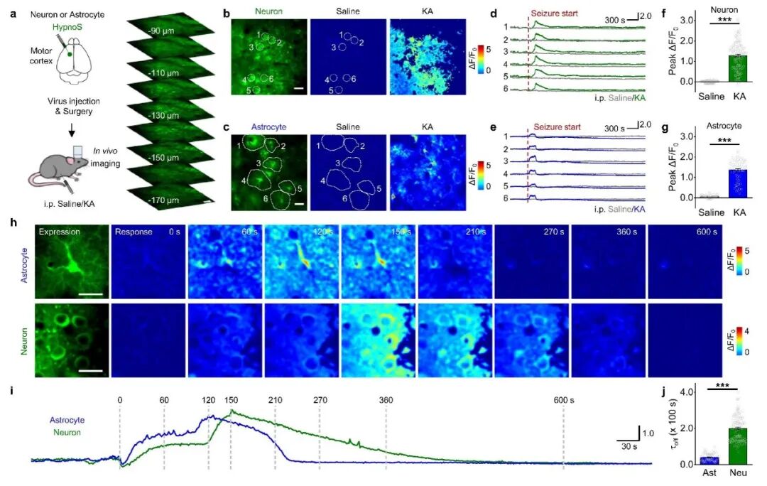
Previous studies have shown that iAdo exerts a protective effect against epileptic seizures in various brain regions, including the hippocampus and cortex. Using the novel HypnoS fluorescent sensor together with large-field-of-view in vivo imaging, the authors were the first to achieve dynamic monitoring of adenosine across the whole cerebral cortex of living mice during KA-induced seizures. In addition, by expressing HypnoS under neuron-specific or astrocyte-specific conditions, the authors investigated the differences in iAdo dynamics between neurons and astrocytes. They found that neuronal and astrocytic HypnoS both showed significant fluorescence increases after KA injection, and that the Ado rise in neurons exhibited a longer decay time than in astrocytes. This implies different iAdo metabolic pathways in neurons and astrocytes in vivo. These results demonstrate the ability of HypnoS to report iAdo dynamics in mice during seizure activity with high sensitivity and with molecular and cell-type specificity.
Figure 2 In vivo two-photon imaging resolves cell-type-specific iAdo dynamics
3. ENT1/2 regulate iAdo levels across the sleep–wake cycle
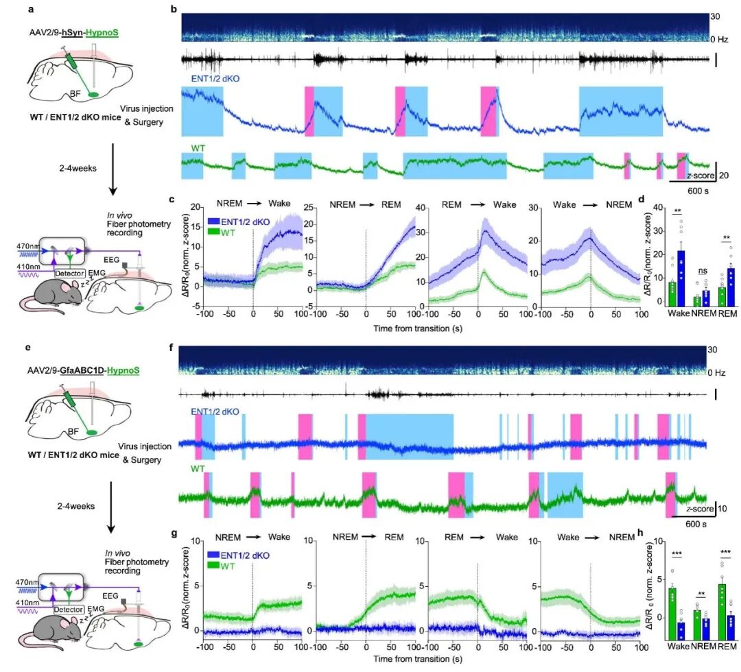
Equilibrative nucleoside transporters (ENT1/2) are essential for regulating iAdo levels and their coupling to eAdo. Neuronal iAdo dynamics during different sleep–wake states largely reflected the neuronal activity-related eAdo dynamics recorded by the separated sensor GRABAdo, indicating tight coupling between iAdo and eAdo. Under conditions of glycolytic inhibition and neuronal activation, iAdo levels were significantly increased in ENT1/2 knockout neurons compared with WT neurons. Conversely, in previous studies of ENT1/2 KO neurons, the authors detected no increase in eAdo after using the ENT1/2 inhibitor S-250. The authors further compared iAdo dynamics recorded by HypnoS during the sleep–wake cycle with iAdo dynamics in the BF of wild-type mice, and observed that ENT1/2 dKO significantly increased iAdo in neurons during wakefulness and REM sleep, whereas in astrocytes iAdo was significantly reduced during wakefulness and REM sleep. These results demonstrate the ability of HypnoS to detect cell-type-specific ENT1/2-dependent Ado transport in mice.
Figure 3 HypnoS reveals ENT1/2-mediated adenosine transport across the sleep–wake cycle
Conclusions
This study developed HypnoS, a brand-new fluorescent sensor for intracellular adenosine, explored the cell-type-specific spatiotemporal dynamics of intracellular adenosine during physiological and pathological processes in living animals, and visualized ENT1/2-mediated adenosine transmembrane transport in neurons and astrocytes, providing a new tool for understanding the mechanisms of action of adenosine.