CELL DEATH DIS: Hongmei Yang's team at Huazhong University of Science and Technology reveals the core molecular signalling axis of muscle wasting in lung cancer cachexia
AAV9-PEBP4 Virus Packaging Case | Hongmei Yang's team at Huazhong University of Science and Technology reveals in Cell Death & Disease (IF 12.2) that the PEBP4-NRF2 axis alleviates muscle wasting in lung cancer cachexia
Paper title: PEBP4 alleviates muscle wasting in lung cancer cachexia via KEAP1–NRF2-mediated redox homeostasis
Journal: Cell Death & Disease (IF 12.2)
Collaborating partner: School of Basic Medicine, Tongji Medical College, Huazhong University of Science and Technology (Hongmei Yang team)
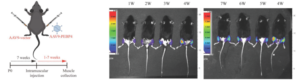
In vivo fluorescence imaging confirms stable expression of AAV9-PEBP4 in GA muscle from 1 to 7 weeks after injection
Research Background
Cancer-associated cachexia (CAC) is a multifactorial metabolic syndrome characterised by progressive skeletal muscle wasting. However, the molecular link between tumour metabolic stress and muscle degeneration remains unclear. On 3 June 2026, the team of Professor Hongmei Yang at the School of Basic Medicine, Tongji Medical College, Huazhong University of Science and Technology, published in Cell Death & Disease (IF 12.2) the paper entitled PEBP4 alleviates muscle wasting in lung cancer cachexia via KEAP1–NRF2-mediated redox homeostasis, which reveals that the tumour lactate-PEBP4-NRF2 axis links cancer metabolism with redox imbalance and muscle wasting, and indicates the therapeutic potential of targeting the PEBP4-NRF2 pathway for lung cancer-associated cachexia.
Research Results
1. Mechanism by which PEBP4 alleviates oxidative stress in skeletal muscle in vitro and in vivo
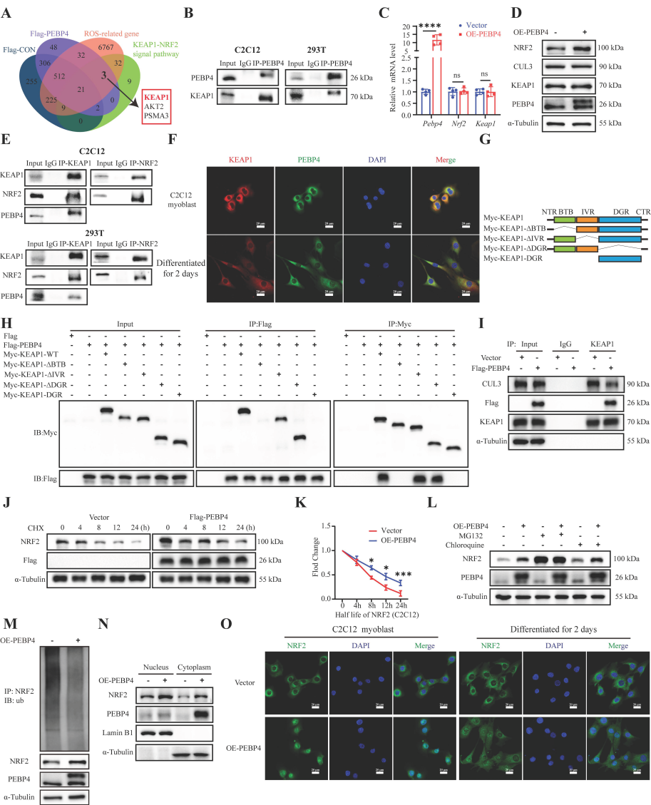
First, the authors identified key regulators associated with CAC-induced muscle wasting, and through RNA-seq data and in vitro and in vivo experimental validation showed that PEBP4 is a key gene in CAC muscle wasting; tumour-derived lactate promoted the enrichment of H3K18la on the Pebp4 promoter, thereby inhibiting Pebp4 transcription in skeletal muscle cells. In addition, PEBP4 functionally regulates the expression of atrophy-related genes and maintains the integrity of muscle structure under physiological conditions. Next, the authors explored the molecular mechanism of PEBP4-mediated muscle protection and found that, under lung CAC conditions, PEBP4 rebuilds redox homeostasis through the NRF2-mediated antioxidant pathway, thereby protecting skeletal muscle from oxidative damage. Further study found that PEBP4 stabilises NRF2 by disrupting Keap1-CUL3-mediated ubiquitination, thereby promoting its nuclear accumulation and supporting the transduction of NRF2-driven antioxidant signalling under lung cancer cachexia stress.
Figure 1. PEBP4 promotes NRF2 nuclear translocation and antioxidant signalling by disrupting Keap1-mediated ubiquitination
2. AAV9-mediated PEBP4 transduction alleviates muscle wasting and oxidative stress in a mouse lung CAC model
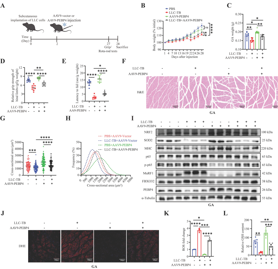
To evaluate the in vivo therapeutic effect of PEBP4, AAV9-PEBP4 was injected into the GA muscle of LLC-TB mice. PEBP4 gene overexpression significantly alleviated CAC-induced body weight loss, preserved skeletal muscle mass and enhanced functional performance. Histological examination showed increased myofibre cross-sectional area and fibre size in PEBP4-treated LLC-TB mice. Western blot analysis showed enhanced activation of the NRF2-SOD2 antioxidant axis, suppressed NF-κB signalling (p-p65), reduced expression of the muscle atrophy markers MuRF1 and FBXO32, and maintained MHC protein levels. AAV9-PEBP4 injection also significantly alleviated oxidative stress in the GA muscle. These results indicate that AAV9-mediated PEBP4 overexpression protects against lung CAC-induced muscle degeneration by strengthening antioxidant defence and inhibiting catabolic remodelling processes.
Figure 2. PEBP4 overexpression improves muscle wasting and ROS accumulation in lung CAC
Conclusions
This study establishes PEBP4 as a redox-regulating effector that links tumour metabolic stress with skeletal muscle degeneration. It reveals a non-canonical mechanism in which PEBP4 disrupts the assembly of the KEAP1–CUL3 complex at the BTB domain, thereby stabilising NRF2 without affecting the binding of KEAP1 to NRF2. This mechanistic distinction highlights the therapeutic potential of targeting the tumour-derived lactate-PEBP4-Keap1-NRF2 axis in cancer-associated muscle wasting and supports the further development of gene- and pharmacology-based clinical therapeutic strategies.