IF 19.1! Jing Zhang and Guoping Peng teams at the First Affiliated Hospital of Zhejiang University publish in TRANSL NEURODEGENER: HSPA8 may become a new target for Alzheimer's disease intervention
AAV_miR30_shRNA Interference Virus Packaging Case | Jing Zhang and Guoping Peng teams at the First Affiliated Hospital, Zhejiang University School of Medicine, reveal in Translational Neurodegeneration (IF 19.1) that HSPA8 regulates the SNARE complex to drive p-tau217+ EV spread
Paper title: HSPA8 orchestrates SNARE complex assembly to drive extracellular vesicle-mediated spread of p-tau217 in Alzheimer’s disease
In patients with Alzheimer's disease (AD), free p-tau217 in plasma is elevated, yet the proportion of p-tau217-positive extracellular vesicles (EVs) is reduced — this paradoxical phenomenon suggests an abnormal regulatory mechanism of EVs in AD. In August 2026, the team of Professors Jing Zhang and Guoping Peng at the First Affiliated Hospital, Zhejiang University School of Medicine, published in Translational Neurodegeneration (IF 19.1) a study entitled HSPA8 orchestrates SNARE complex assembly to drive extracellular vesicle-mediated spread of p-tau217 in Alzheimer’s disease, revealing the role of HSPA8 in regulating multivesicular body (MVB)-mediated EV release and tau protein spreading, and highlighting HSPA8 as a promising therapeutic target for altering AD progression.
Research Results
1. Reduced synthesis/secretion of p-tau217+EVs
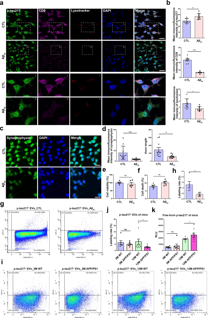
The team confirmed that free p-tau217 in the cerebrospinal fluid and plasma of patients with AD was significantly higher than in healthy controls, reproducing the paradoxical phenomenon of “elevated free p-tau217 with decreased p-tau217+EVs”. They then constructed an in vitro three-cell co-culture blood-brain barrier (BBB) model and found that in the BBB damaged by Aβ42 treatment, the proportion of EVs crossing the barrier was actually higher, suggesting that the reduction of p-tau217+EVs in peripheral blood is not caused by impaired trans-BBB transport of EVs from the central nervous system to the periphery. After SH-SY5Y cells were treated with Aβ42, intracellular p-tau217 accumulated; lysosomal degradation capacity decreased, but the impairment of p-tau217+EV synthesis and secretion was even more pronounced, suggesting that a secretion defect, rather than abnormal degradation, is the main cause of the vesicle reduction. In addition, Aβ42 treatment did not cause a significant decrease in cell viability or cytotoxicity, but did induce synaptic damage. Nano-flow cytometry of the cell supernatant confirmed a marked decrease in p-tau217+EV secretion. By analysing multiple phosphorylation sites in Aβ42-treated SH-SY5Y cells, they demonstrated that it is the selective vesicular packaging and secretion of a specific phosphorylated tau isoform that is impaired, rather than a change in total tau expression. In in vivo validation in APP/PS1 mice, plasma free p-tau217 was elevated and the proportion of p-tau217+EVs was significantly reduced in 12-month-old aged AD mice; 3-month-old young mice did not yet show these changes, consistent with the age-dependent pathological features of AD. Together, these in vitro and in vivo experiments indicate that the synthesis and secretion capacity of neuronal p-tau217+EVs is impaired, leading to a reduction of peripheral p-tau217+EVs and intracellular accumulation of p-tau217.
Levels of p-tau217 and p-tau217+EVs in Aβ42-treated cells or APP/PS1 mice
2. HSPA8 regulates SNARE complex assembly and p-tau217+EV secretion
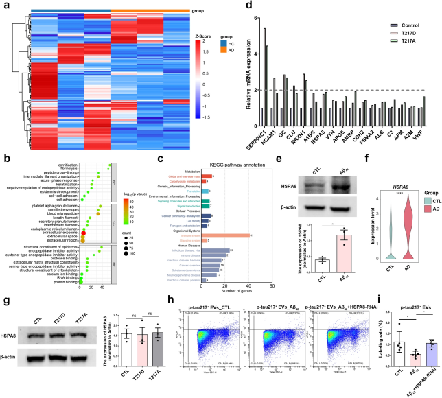
The team performed mass spectrometry-based proteomic analysis of p-tau217+EVs immunocaptured from the cerebrospinal fluid of patients with AD, and identified core hub genes through GO, KEGG and PPI interaction network screening. Using tau-T217 phosphorylation/dephosphorylation mimic mutants to distinguish upstream and downstream molecules, they found that several genes, including HSPA8, were not regulated by the phosphorylation state of p-tau217 and may act upstream. Among these genes, HSPA8 has been reported to regulate SNARE complex function and to mediate vesicle synthesis and secretion. The team found that HSPA8 protein expression was upregulated in Aβ42-treated SH-SY5Y cells; human hippocampal spatial transcriptomic data confirmed that HSPA8 gene expression was also elevated in the hippocampus of patients with AD. They constructed an HSPA8 shRNA knockdown model, and nano-flow cytometry results showed that Aβ42 treatment caused a decrease in p-tau217+EV secretion, while HSPA8 knockdown significantly restored the secretion level of p-tau217+EVs, confirming that HSPA8 negatively regulates p-tau217+EV release. Further mechanistic exploration revealed that abnormally elevated HSPA8 in AD impairs p-tau217+EV secretion by promoting SNAP29-Aβ binding and disrupting SNARE complex assembly.
HSPA8 is an upstream signalling molecule regulating p-tau217-associated EVs
3. HSPA8 inhibition improves cognition and restores p-tau217+EV secretion
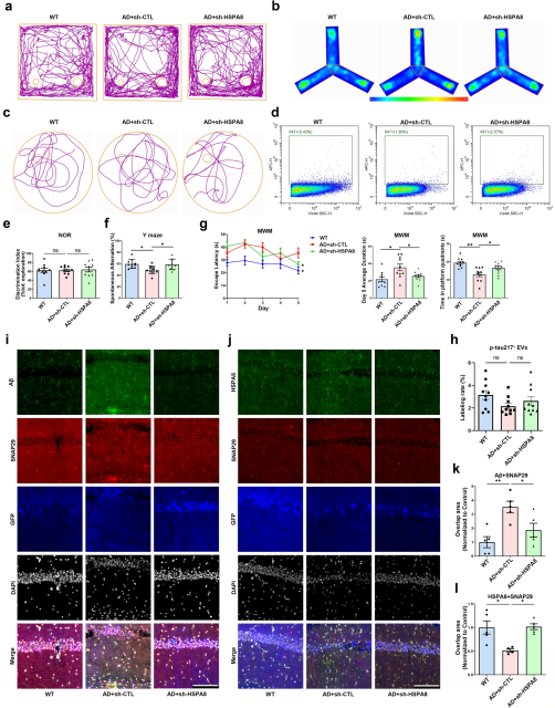
To further investigate the role of HSPA8 in p-tau217+EV synthesis/secretion and AD progression, the team stereotaxically injected AAV-shHSPA8 into the hippocampus of 6-month-old APP/PS1 mice, and found that HSPA8 knockdown alleviated the cognitive deficits of APP/PS1 mice. Nano-flow cytometry of mouse plasma showed a decreasing trend in the proportion of p-tau217+EVs in APP/PS1 mice, and hippocampal HSPA8 knockdown reversed this decrease and restored plasma p-tau217+EV levels. Immunofluorescence of hippocampal tissue showed that, compared with the control group, the sh-HSPA8 group had significantly reduced Aβ-SNAP29 co-localisation and restored HSPA8-SNAP29 co-localisation, reproducing the in vitro molecular mechanism in vivo. These findings indicate that HSPA8 inhibition not only improves cognitive dysfunction in APP/PS1 mice but also restores the synthesis and secretion of p-tau217+EVs. Based on the binding site between HSPA8 and SNAP29, the team designed and synthesised a SNAP29-derived 25-mer competitive inhibitory peptide to block the abnormal HSPA8-mediated Aβ-SNAP29 interaction, and confirmed that the HSPA8-targeting 25-mer peptide can competitively block abnormal Aβ-SNAP29 binding, restore p-tau217+EV secretion, alleviate tau pathology and improve cognitive deficits in AD model mice, showing potential for AD intervention.
HSPA8 knockdown restores cognitive function and p-tau217+EV secretion in APP/PS1 mice
Conclusions
This study identifies HSPA8 as a key regulator of neuronal p-tau217+EV secretion and reveals its abnormal elevation in AD neurons. Upregulated HSPA8 impairs SNARE complex formation by altering the SNAP29–Aβ interaction, reducing p-tau217+EV release and aggravating AD pathology. Targeting HSPA8 restores EV secretion and improves cognition, providing a promising new avenue for AD intervention.