Neuron | Yidong Shen, Xiumin Yan and Yifei Zhou research teams find that inhibiting macrophage-derived CTSS reverses age-related brain barrier damage
AAV2/5 intracerebroventricular injection for CTSS study of the ageing blood-CSF barrier | Yidong Shen and Yifei Zhou with Xiumin Yan, Neuron (IF 14.7)
Paper title: Macrophage-derived CTSS drives the age-dependent disruption of the blood-CSF barrier
Journal: Neuron (IF 14.7)
Collaborating partner: Center for Excellence in Molecular Cell Science, Chinese Academy of Sciences - Yidong Shen and Yifei Zhou research group, in collaboration with Xinhua Hospital Affiliated to Shanghai Jiao Tong University School of Medicine - Xiumin Yan group
· With WZBIO Support - AAV ·
Gene information
CTSS: cathepsin S
HRAS(G12V): oncogene
CLDN1: claudin-1
Cystatin C: a secreted protein and a potent endogenous inhibitor of CTSS
The choroid plexus (CP) is the main source of cerebrospinal fluid (CSF), and the blood-CSF barrier is formed by tight junctions between epithelial cells in the CP and is essential for maintaining brain homeostasis. Dysfunction of this barrier is associated with brain ageing and neurodegenerative diseases, but the mechanism of its degeneration during ageing remains unclear. On 19 February 2025, the research group of Yidong Shen and Yifei Zhou at the Center for Excellence in Molecular Cell Science (Shanghai Institute of Biochemistry and Cell Biology), Chinese Academy of Sciences, in collaboration with the group of Xiumin Yan at Xinhua Hospital Affiliated to Shanghai Jiao Tong University School of Medicine, published in Neuron (IF 14.7) a paper entitled Macrophage-derived CTSS drives the age-dependent disruption of the blood-CSF barrier. The study revealed an important interaction between immune cells and barrier cells that accelerates ageing of the CP and the brain, identified cathepsin S (CTSS) as a potential target for improving brain homeostasis in aged animals, and highlighted the key role of circulating proteases in ageing.
Research Results
1. The blood-CSF barrier is impaired with age, with a corresponding decrease in CLDN1
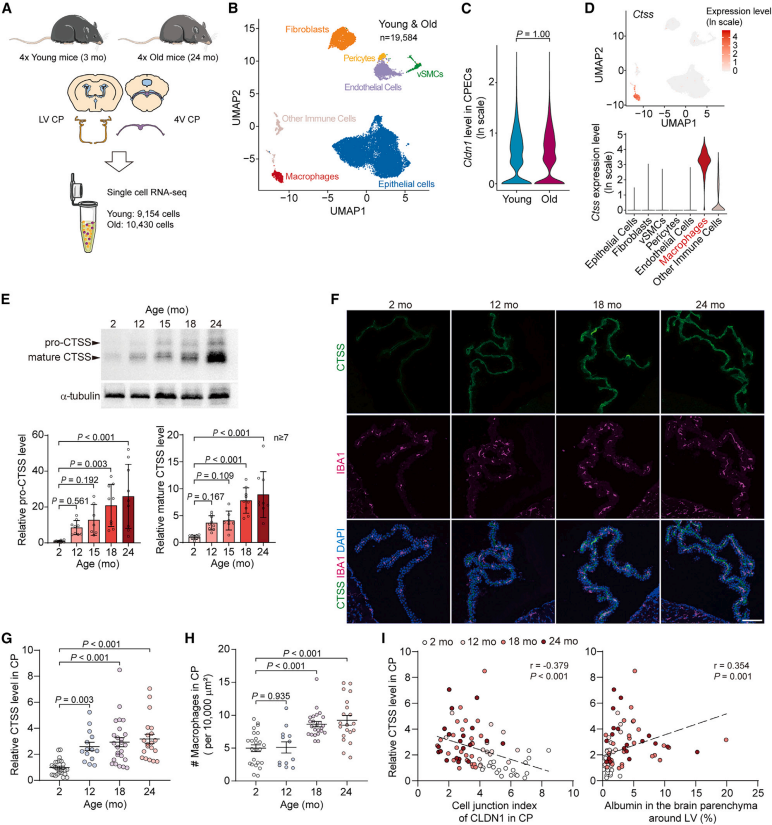
The authors first investigated whether the reduction of CLDN1 at CPEC TJs leads to damage of the B-CSF barrier during ageing. The study found that CLDN1-dependent disruption of the B-CSF barrier in the aged CP may play an important role in age-related neurological dysfunction by promoting T cell infiltration in the subventricular zone (SVZ) and reducing cell proliferation. To explore the mechanism that disrupts CLDN1 at CPEC TJs during ageing, the authors performed single-cell RNA sequencing (scRNA-seq) on the CP of the left ventricle and fourth ventricle of 3-month-old and 24-month-old mice, and tracked the temporal transcriptome of individual CP cells. The results showed that CTSS was highly expressed in macrophages and increased in an age-dependent manner, whereas its expression in other cells showed no obvious change. Further experiments confirmed that the expression and secretion of CTSS increased in the ageing choroid plexus and originated mainly from macrophages. By comparing CTSS levels in the CP with CLDN1 intensity at CPEC TJs and with albumin signals around the left ventricle during ageing, the authors found that both correlated markedly with CTSS levels. In summary, during ageing, CTSS is induced by macrophages and secreted into the CP stroma, permeabilising the B-CSF barrier by digesting CLDN1. Further studies found that the age-dependent increase of CTSS in the CP impairs the B-CSF barrier.
Figure 1. CTSS is upregulated in the ageing choroid plexus
2. CTSS weakens the B-CSF barrier through CLDN1
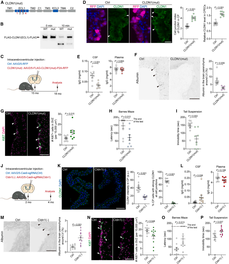
To further investigate whether the CTSS-mediated reduction of CLDN1 is the cause of the age-dependent degradation of the B-CSF barrier, the authors injected AAV2/5 expressing a CLDN1 mutant resistant to CTSS cleavage (CLDN1mut) into the left ventricle of 15-month-old mice. After CLDN1mut overexpression, CLDN1 signal at CPEC TJs increased and the B-CSF barrier improved. CLDN1mut also increased proliferating cells in the SVZ and enhanced the behaviour of mice in the Barnes maze and tail suspension tests. To confirm the key role of CLDN1 in maintaining the B-CSF barrier, the authors downregulated CLDN1 in CPECs of young mice by injecting AAV2/5 expressing Cas9 and an sgRNA targeting CLDN1. CLDN1 downregulation disrupted the B-CSF barrier, inhibited cell proliferation in the SVZ, and impaired mouse behaviour in the Barnes maze and tail suspension tests. Therefore, CTSS-induced reduction of CLDN1 at CPEC TJs is essential for the age-dependent breakdown of the B-CSF barrier.
Figure 2. CTSS weakens the B-CSF barrier through CLDN1
3. Cellular senescence in the aged CP intensifies CTSS induction
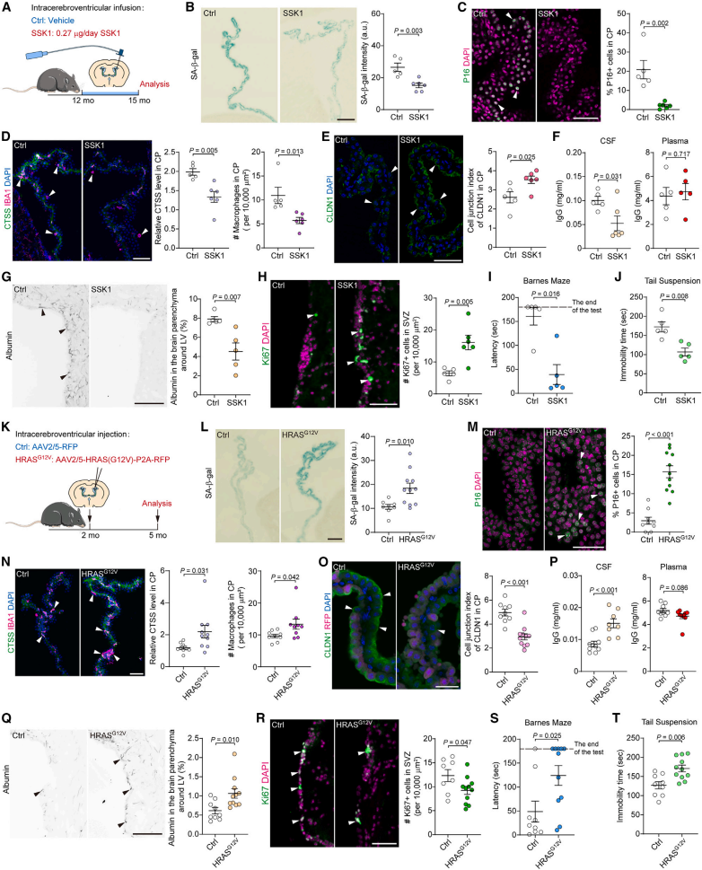
To investigate the mechanism by which stromal macrophages in the aged CP secrete CTSS, the authors examined cellular senescence in the CP. During CP ageing, cellular senescence increased significantly in CPECs, fibroblasts, vSMCs, pericytes and endothelial cells. CCL2 is an inflammatory SASP factor that binds to CCR2 and recruits monocytes, and CCL2 was found to increase in the aged CP. To study the role of CCL2 in CCR2 KO mice, the authors found that CTSS levels in the CP of 12-month-old CCR2 KO mice were significantly lower than those in WT mice. It was further confirmed in vivo that removing senescent cells from the aged CP attenuated CTSS induction and restored the disrupted B-CSF barrier and the associated neurological dysfunction. Oncogenic Ras (G12V) is an established method for inducing cellular senescence, and specific overexpression of Ras (G12V) in CPECs using AAV2/5 induced cellular senescence in the CP of young mice. Ras (G12V)-induced cellular senescence increased macrophages and CTSS in the CP, reduced CLDN1 at CPEC TJs, disrupted the B-CSF barrier, and inhibited cell proliferation in the SVZ, further supporting that senescent cells in the CP drive CTSS-mediated disruption of the B-CSF barrier. Therefore, the age-dependent increase in cellular senescence in the CP most likely activates macrophage CTSS secretion, leading to disruption of the B-CSF barrier.
Figure 3. Interfering with cellular senescence in the CP alters the B-CSF barrier
Conclusions
This study reports that cathepsin S (CTSS), a protease secreted by CP macrophages, is upregulated in the aged CP as a result of exacerbated cellular senescence, and that CTSS impairs the blood-CSF barrier by digesting CLDN1. Notably, inhibiting CTSS or upregulating CLDN1 in the aged CP restores the blood-CSF barrier and brain function.