Neuron | Overturning conventional understanding: the new role of non-image-forming photoreceptors ipRGCs in vision
AAV2/2-DIO-GFP Custom | USTC Tian Xue and Zhong Li teams reveal that non-image-forming photoreceptors ipRGCs improve visual orientation selectivity and image perception (Neuron, IF 14.7)
Paper title: Non-image-forming photoreceptors improve visual orientation selectivity and image perception
Journal: Neuron (IF 14.7)
Collaborating partner: University of Science and Technology of China (Tian Xue, Zhong Li and Rong Liu teams), in collaboration with Army Medical University (Xiaowei Chen team), the Shenzhen Institute of Advanced Technology (Jin Bao team) and Tsinghua University (Yuanyuan Mi team)
For a long time, image perception was thought to be mediated solely by rods and cones, with intrinsically photosensitive retinal ganglion cells (ipRGCs) responsible only for non-image-forming visual functions. However, earlier studies hinted that ipRGCs might play a potential role in contrast detection, and whether they influence cortical visual feature processing and image recognition in mammals remained unclear. Recently, the teams of Tian Xue, Zhong Li and Rong Liu of the Division of Life Sciences and Medicine, University of Science and Technology of China, together with Professor Xiaowei Chen's team of Army Medical University, Professor Jin Bao's team of the Shenzhen Institute of Advanced Technology and Professor Yuanyuan Mi's team of Tsinghua University, published online in Neuron (IF 14.7) the paper Non-image-forming photoreceptors improve visual orientation selectivity and image perception. The study found that ipRGCs can improve visual orientation selectivity and image perception by regulating excitatory and inhibitory neurons in the primary visual cortex (V1), revising the decades-old view that image contour perception is mediated solely by rods and cones.
Research Results
1. ipRGCs enhance the orientation selectivity of V1 neurons
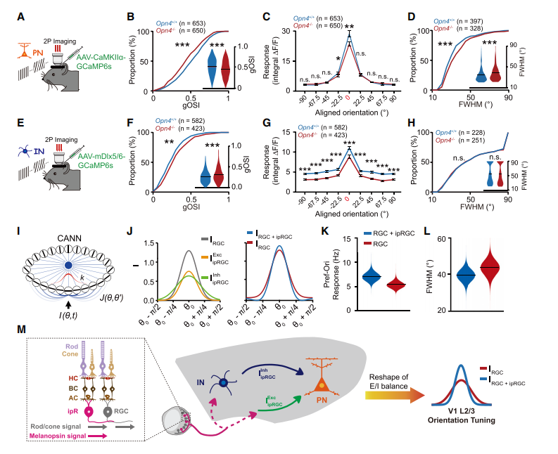
Using in vivo two-photon calcium imaging, the researchers recorded the responses of V1 L2/3 neurons loaded with the calcium indicator Cal-520 AM in mice to drifting grating visual stimuli at 16 orientations. V1 orientation selectivity was characterized by measuring the global orientation selectivity index (gOSI) and the tuning bandwidth (FWHM). Comparing Opn4-/- mice (in which deficiency of the Opn4 gene prevents melanopsin expression and ipRGCs lose their intrinsic photosensitivity) with wild-type mice, they found that V1 L2/3 neurons in Opn4-/- mice showed a significantly reduced gOSI, a broader FWHM and weaker Ca2+ responses to the preferred orientation (Pref-Ori). Subsequent melanopsin expression-restoration and interference experiments fully demonstrated that melanopsin expression is essential for improving V1 orientation selectivity. Using chemogenetic tools to lower ipRGC activity and to alter the visual spectrum further confirmed that activation of ipRGCs is crucial for enhancing orientation selectivity in the visual cortex. In-depth analysis showed that ipRGCs can independently activate both excitatory and inhibitory neurons in V1 L2/3, and that they can transmit melanopsin-dependent light signals to V1 cortical neurons, thereby exciting both types of neurons. Finally, the team explored the regulatory properties of ipRGCs on V1 excitatory neurons and inhibitory interneurons. The results showed that ipRGC activation has differential effects on V1 excitatory and inhibitory neurons, and that this effect reshapes the cortical excitatory–inhibitory balance, thereby enhancing V1 orientation selectivity.
Light-activated ipRGCs reshape the cortical excitatory–inhibitory balance to improve orientation selectivity
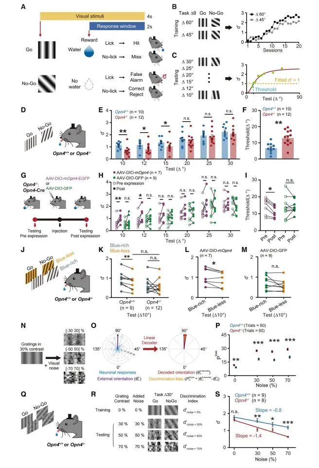
2. Light-activated ipRGCs improve the orientation discrimination ability of mice behaviourally
To investigate the effect of ipRGCs on the behaviour of image visual perception, the team subjected mice to a series of Go/No-Go tasks to test their orientation discrimination ability. In the task with small orientation differences, wild-type mice showed a significantly larger d' value than Opn4-/- mice and a lower orientation discrimination threshold, indicating that wild-type mice perform better at discriminating orientations with small angular differences, that is, they have a stronger orientation discrimination ability. Restoring melanopsin expression in the ipRGCs of Opn4-/-;Opn4-Cre mice significantly improved the orientation discrimination ability of these mice, whereas mice injected with GFP as a control showed no obvious change in orientation discrimination ability. These results indicate that melanopsin expression in ipRGCs is essential for improving the orientation discrimination ability of mice. Testing with “blue-rich” and “blue-poor” gratings in a Δ10° task further confirmed that photoactivation of ipRGCs improves the orientation discrimination ability of mice. In addition, consistent with the animal experiments, photoactivation of ipRGCs also improved visual orientation discrimination in humans. By studying the effect of cortical input from ipRGCs on resistance to external visual noise, the team found that cortical input from ipRGCs can enhance the resistance of V1 to external visual noise, which is of great significance for understanding how the visual system processes information in complex environments.
Photoactivation of ipRGCs improves orientation discrimination in mice
Conclusions
This study reveals that ipRGCs improve visual feature processing by reshaping the cortical excitatory–inhibitory balance, thereby enhancing visual orientation discrimination in both mice and humans. This finding revises the decades-old traditional view of image contour perception and highlights the importance of the melanopsin-sensitive spectrum in optimizing light conditions for work and daily life.