IF 14.1: Why Is Pancreatic Cancer "Unmoved" by PD-1? Prof. Gang Zhao's Team at Union Hospital, Tongji Medical College, HUST Reveals MCT1-Mediated Lactate Shuttling as the Key
Paper title: Targeting Lactate-Driven Stromal Autophagy via MCT1 Disrupts the Immunosuppressive Niche and Sensitizes Pancreatic Cancer to PD-1 Blockade
Journal: Advanced Science (IF 14.1)
Collaborating partner: Prof. Gang Zhao's team, Union Hospital, Tongji Medical College, Huazhong University of Science and Technology
Lactate reshapes the tumor microenvironment (TME) through complex communication between cancer and stromal cells, and pancreatic stellate cells (PSCs) are a key TME component that drives tumorigenesis; however, whether lactate mediates the crosstalk between pancreatic cancer (PC) cells and PSCs remains unclear. In May 2026, Prof. Gang Zhao's team at Union Hospital, Tongji Medical College, Huazhong University of Science and Technology published in Advanced Science (IF 14.1) an article entitled Targeting Lactate-Driven Stromal Autophagy via MCT1 Disrupts the Immunosuppressive Niche and Sensitizes Pancreatic Cancer to PD-1 Blockade, which identified lactate as a key TME signal molecule that orchestrates PSC activation and the immunosuppressive microenvironment, providing evidence for the combined use of MCT1 inhibition and immune checkpoint blockade in the treatment of pancreatic cancer.
Research Findings
1. Lactate induces autophagy in PSCs by regulating Vps34 lactylation
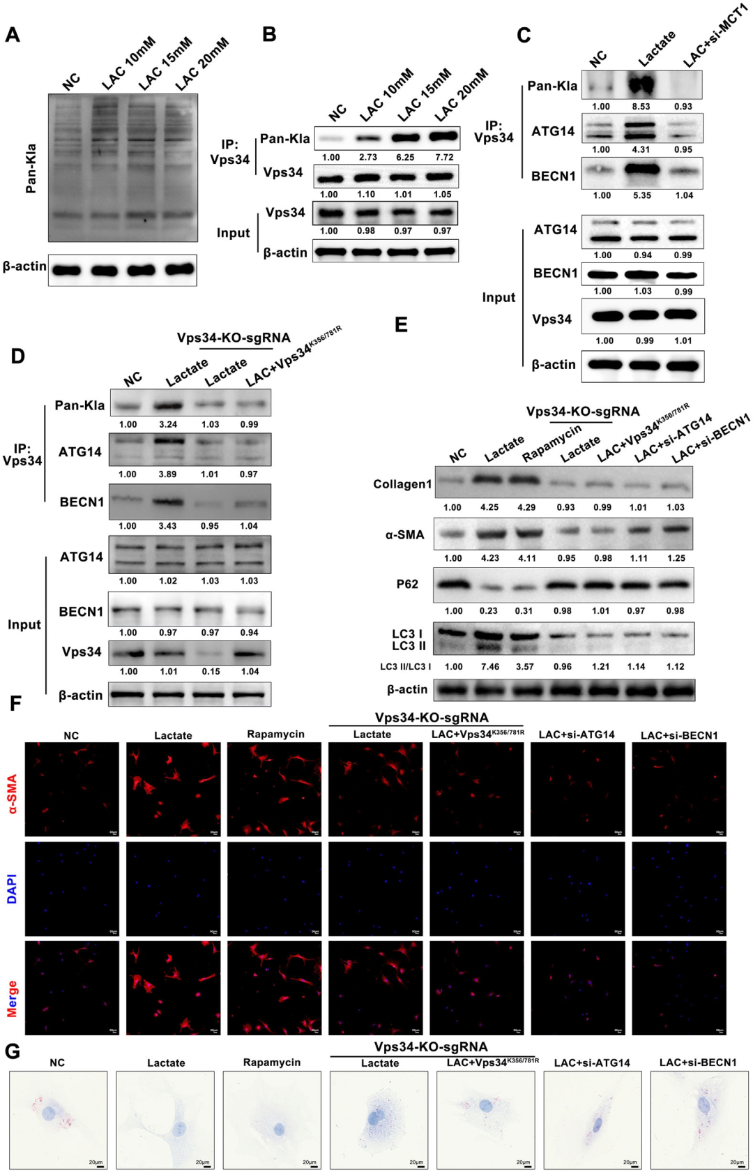
The authors found that lactate is an important signaling molecule mediating the crosstalk between PC cells and PSCs. Lactate is mainly produced by PC cells and can be extensively shuttled to PSCs, thereby mediating PSC activation. Mechanistically, lactate enters PSCs mainly through the MCT1 transporter on the cell membrane and then activates PSCs in an autophagy-dependent manner. Vps34 promotes autophagosome formation, and the authors further explored whether lactate induces autophagy in PSCs through lactylation of Vps34. Pan-lactylation antibody Western blot analysis showed that lactate increases the overall lactylation level in PSCs, and Co-IP experiments showed that lactate significantly increases the lactylation level of Vps34 and promotes the recruitment of Vps34 to ATG14 and BECN1; however, this effect was significantly impaired in Vps34-knockout PSCs or in PSCs overexpressing Vps34 K356/781R after Vps34 knockout. In addition, Western blot, α-SMA IF, collagen I ELISA and other experiments also showed that lactate-induced autophagy and activation of PSCs were markedly reversed in Vps34-knockout PSCs or in PSCs overexpressing Vps34 K356/781R after Vps34 knockout. These results demonstrate the importance of Vps34 lactylation in lactate-induced autophagy initiation and activation of PSCs.
Figure 1. Lactate induces autophagy in PSCs by regulating Vps34 lactylation
2. Orthotopic pancreatic cancer development is impaired in PSCs-MCT1−/− mice
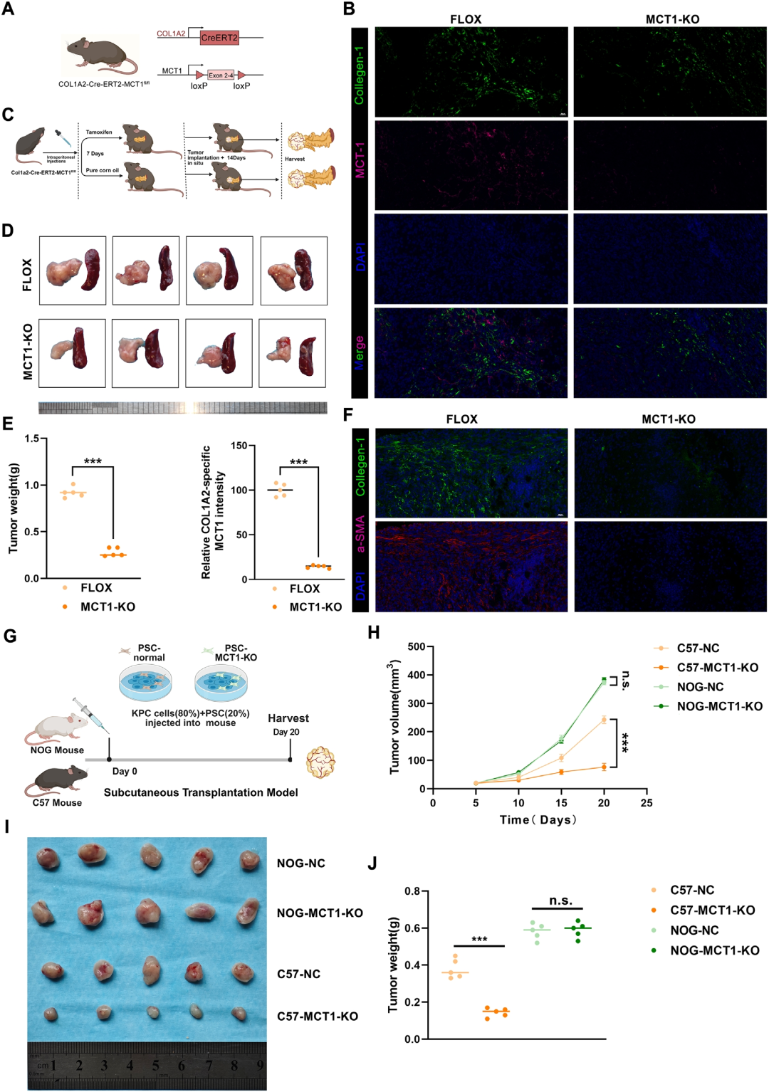
To further reveal the effect of lactate-activated PSCs on PC in vivo, the authors generated PSC-specific MCT1 knockout mice (PSCs-MCT1−/−), and then injected KPC cells orthotopically into the pancreas of knockout and control mice to establish orthotopic tumor models. The tumors of PSCs-MCT1−/− mice were significantly smaller and lighter than those of control mice, and PSC activation was significantly reduced. In vivo experiments showed that MCT1−/− mice could significantly slow tumor growth and inhibit liver metastasis of PC, which was consistent with the effect of PSCs on the invasion and migration of PC cells in in vitro co-culture assays, but inconsistent with the effect of PSCs on the proliferative capacity of PC. Therefore, the authors hypothesized that activated PSCs may not directly enhance tumor proliferation, but rather indirectly enhance tumor proliferation by modulating the immune TME. The authors established subcutaneous KPC and PSC co-xenograft tumor models in immunocompetent C57 mice or immunodeficient NOG mice, and found that the growth of co-transplanted tumors with MCT1-knockdown PSCs was significantly inhibited in C57 mice but not in NOG mice, indicating that lactate-activated PSCs mainly promote PC tumorigenesis by manipulating the immune TME.
Figure 2. Orthotopic pancreatic cancer development is suppressed in PSCs-MCT1−/− mice
3. AZD3965 sensitizes orthotopic pancreatic cancer to PD-1 blockade to suppress tumor progression in mice
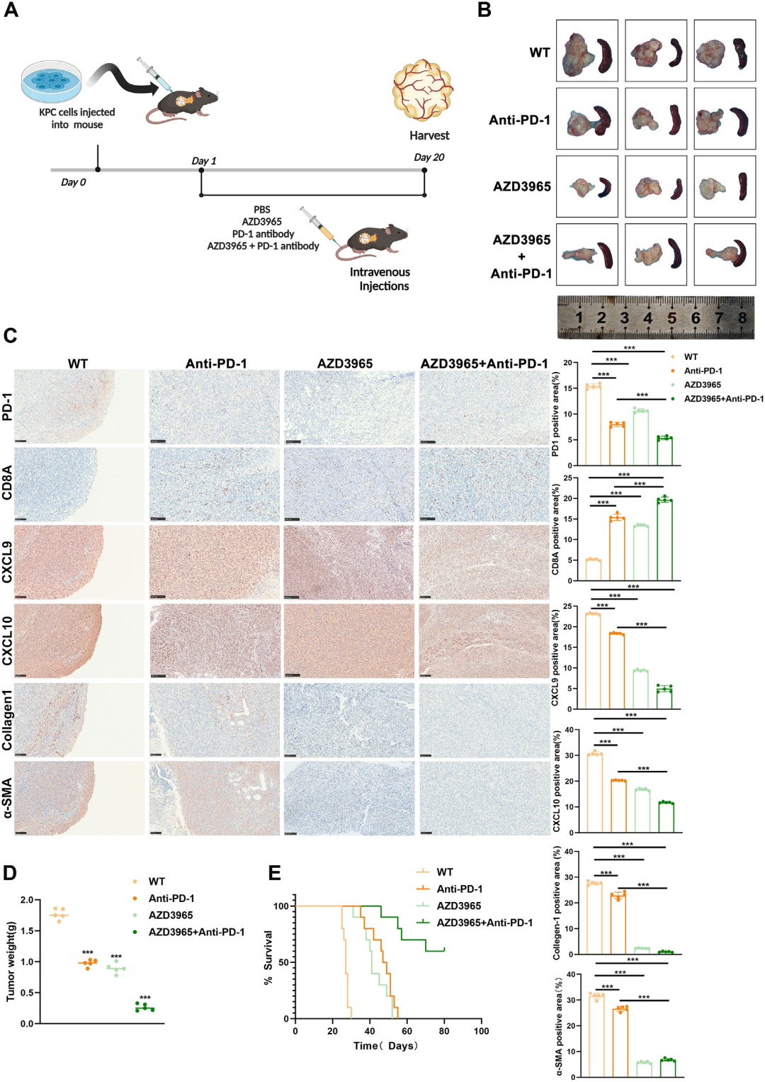
Mechanistic studies revealed that lactate-activated PSCs upregulate PD-1 expression in CD8+ T cells through the CXCL9/CXCL10-CXCR3-STAT3 axis, thereby inducing an immunosuppressive TME. The authors investigated whether the MCT1-specific inhibitor AZD3965 could sensitize PC to anti-PD-1 antibody therapy, thereby suppressing tumor growth. KPC cells were orthotopically injected into the pancreas of mice, which were then treated with PBS, AZD3965, anti-mouse PD-1 antibody, or the combination of AZD3965 and anti-mouse PD-1 antibody. The results showed that AZD3965 or anti-mouse PD-1 antibody monotherapy significantly inhibited tumor growth, but the combination of AZD3965 and anti-mouse PD-1 antibody further inhibited tumor growth. Compared with the control group, anti-mouse PD-1 antibody therapy reduced the PD-1-positive area but increased CD8+ T cell infiltration. AZD3965 alone significantly increased CD8+ T cell infiltration and reduced tissue fibrosis and CXCL9/CXCL10 levels, and this effect was further enhanced in combination with anti-mouse PD-1 antibody. In addition, AZD3965 sensitized tumors to anti-mouse PD-1 antibody, significantly suppressing tumor growth and improving the survival of PC mice. These results indicate that AZD3965 sensitizes tumors to anti-mouse PD-1 antibody and effectively suppresses PC progression by modulating fibrosis and the immunosuppressive TME.
Figure 3. AZD3965 sensitizes orthotopic pancreatic cancer to PD-1 blockade to suppress tumor progression in mice
Conclusions
This study reveals the critical role of lactate in regulating immune evasion responses and promoting PC progression through MCT1-mediated PSC activation of autophagy, which may provide new insights into the causes of the immunosuppressive microenvironment in PC and potential therapeutic interventions.