Nanjing Medical University Ding Ying / Sun Peng / Su Dongming Research Team Reveals That REG3A Secreted by Peritumoral Acinar Cells Accelerates Pancreatic Cancer Progression
Custom Adenovirus Ad-REG3A | Nanjing Medical University Ding Ying Team Reveals How REG3A Drives Pancreatic Ductal Adenocarcinoma Progression (Cell Communication and Signaling, IF 8.2)
Paper title: REG3A secreted by peritumoral acinar cells enhances pancreatic ductal adenocarcinoma progression via activation of EGFR signaling
Journal: Cell Communication and Signaling (IF 8.2)
Collaborating partner: Nanjing Medical University, Prof. Ding Ying, Sun Peng and Su Dongming
Human pancreatic cancer cell lines SW1990, PANC-1, AsPC-1 and BxPC-3
Viral dose
109
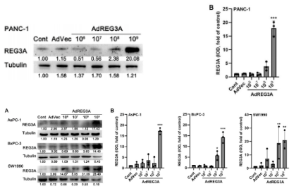
Western blot detection of Ad-REG3A overexpression efficiency in SW1990, PANC-1, AsPC-1 and BxPC-3 cells
Research Background
Pancreatic ductal adenocarcinoma (PDAC) is a lethal cancer with a five-year survival rate of less than 10%, no effective means of early diagnosis and an extremely poor prognosis. REG3A plays a role in the development of a range of malignancies, including PDAC, but because its tissue localisation, receptor and downstream signal transduction mechanisms remain unclear, the role of REG3A in PDAC is still controversial. On 18 February 2025, the research team of Ding Ying, Sun Peng and Su Dongming at Nanjing Medical University published a paper entitled REG3A secreted by peritumoral acinar cells enhances pancreatic ductal adenocarcinoma progression via activation of EGFR signaling in Cell Communication and Signaling (IF 8.2), confirming that increased REG3A expression in peritumoral acinar cells is a specific event indicating PDAC progression and providing a new direction for the diagnosis and treatment of PDAC.
Research Results
1. REG3A promotes PDAC tumour growth in vitro and in vivo
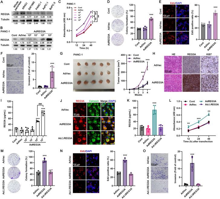
Through a variety of experiments and data analyses, the team found that REG3A expression in peritumoral tissue (rather than in the tumour tissue itself) correlated with malignant progression and poor prognosis in patients with PDAC. To further investigate the effect of REG3A on cultured PDAC cell lines, they showed that in several pancreatic cancer cell lines REG3A expression was lower than in normal pancreatic tissue and in rat AR42J cells. After overexpressing REG3A in the cells using adenovirus, cell viability, colony formation, proliferation and invasion were all enhanced. Subcutaneous xenograft models in nude mice established with PANC-1 or AsPC-1 cells showed that adenovirus-mediated REG3A overexpression stimulated tumour growth in nude mice and that the proliferation rate of the tumour tissue was higher. In addition, treatment with Ad-REG3A increased the REG3A concentration in the culture medium in a dose-dependent manner, and REG3A co-localised with an endoplasmic reticulum marker protein; after removal of the signal peptide, the secreted REG3A was markedly reduced, and Ad-ΔREG3A failed to stimulate the growth of PANC-1 tumour cells, indicating that REG3A functions as a typical secreted protein.
REG3A promotes PDAC tumour growth in vitro and in vivo
2. EGFR-MAPK activation mediates the proliferative effect of secreted REG3A on PDAC cells
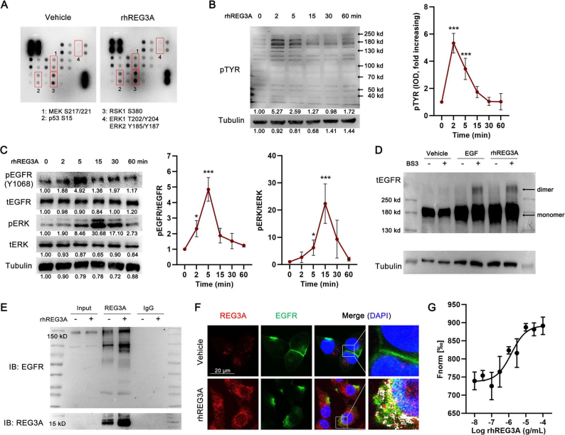
Treatment of PANC-1 and several other PDAC cell lines with recombinant human REG3A (rhREG3A) enhanced cell viability, colony formation, proliferation and invasion, indicating that REG3A promotes tumour growth through paracrine or autocrine models. Transcriptome analysis showed that REG3A stimulation activated oncogenic signalling pathways including EGF/EGFR and MAPK. Treatment of PANC-1 cells with rhREG3A activated the pro-proliferative MAPK pathway, phosphorylating EGFR (Y1068) and downstream ERK; it also induced EGFR dimerisation and co-localised with EGFR, and MST experiments confirmed that the two bind directly. After an EGFR plasmid was transfected into CHO cells that do not originally express EGFR, rhREG3A stimulation activated the EGFR-ERK pathway, whereas the effect on CHO cells transfected with empty vector was small. In silico analysis showed that REG3A binds domains I (L1) and III (L2) of the EGFR extracellular region, similar to the classical binding site of EGF. After deletion of the C-type lectin domain of REG3A, its binding to EGFR was markedly weakened, and REG3A preferentially recognises the extracellular region of EGFR rather than the intracellular kinase domain; EGF competitively inhibited the binding of rhREG3A to EGFR and the downstream phosphorylation of EGFR-ERK.
EGFR-MAPK activation mediates the proliferative effect of REG3A in PDAC cells
3. Inflammation-stimulated STAT3 activation contributes to increased REG3A expression in peritumoral acinar cells
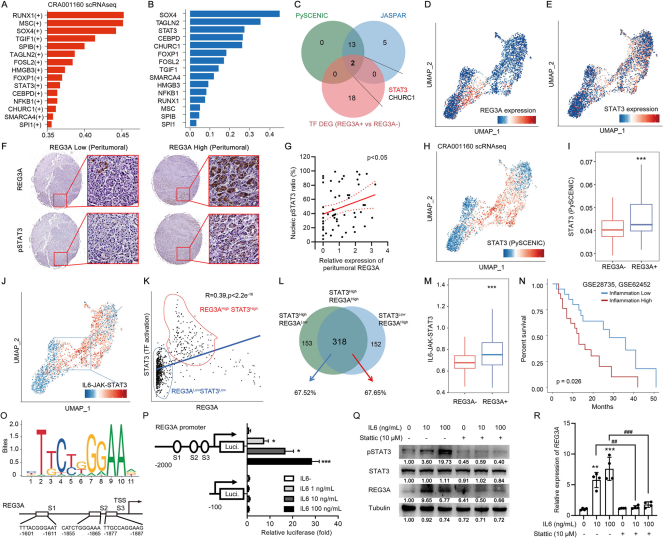
By analysing the transcription factors activated in REG3A-positive acinar cell clusters using PySCENIC, and combining transcription factors upregulated at the mRNA level with their binding to the REG3A gene promoter region, the team found that STAT3 was highly correlated with REG3A upregulation. In the CRA001160 single-cell RNA sequencing dataset, STAT3 mRNA expression was higher in REG3A-positive acinar cells. Immunostaining showed that in the region surrounding PDAC tumours, REG3A expression was significantly positively correlated with phosphorylated STAT3. In addition, both STAT3 activation and enrichment of the IL6-JAK-STAT3 pathway confirmed that activated STAT3 may promote REG3A transcription in acinar cells; activation of the IL6-JAK-STAT3 pathway was associated with a poorer prognosis. In vitro luciferase reporter gene experiments further confirmed that STAT3 can directly transcriptionally activate REG3A. IL6 stimulated STAT3 phosphorylation and increased REG3A expression in a dose-dependent manner, whereas the STAT3 inhibitor Stattic suppressed IL6-induced STAT3 phosphorylation and REG3A mRNA transcription.
Inflammation-induced STAT3 activation upregulates REG3A expression in acinar cells
Conclusions
This study confirms that increased REG3A expression in peritumoral acinar cells indicates PDAC progression, and that REG3A secreted by acinar cells stimulates PDAC cell growth by binding EGFR and activating downstream MAPK signalling. It reveals that REG3A has the potential to become a serum and pathological marker for the diagnosis of PDAC, providing a theoretical basis for the development of new therapeutic approaches.