IF 10.4 | Zhifei Xu's team at the Innovation Institute for Artificial Intelligence in Medicine of Zhejiang University reveals the molecular mechanism of ceritinib-induced cardiotoxicity
Ad-mCherry-GFP-LC3 Adenovirus | Ceritinib Cardiotoxicity Arises from AKT Suppression-Induced Autophagic Flux Impairment: Zhifei Xu's team at Zhejiang University, Acta Pharmacologica Sinica (IF 10.4)
Paper title: AKT (Ser473) suppression mediates ceritinib cardiotoxicity through autophagic flux impairment and mitochondrial injury
Journal: Acta Pharmacologica Sinica (IF 10.4)
Collaborating partner: Innovation Institute for Artificial Intelligence in Medicine of Zhejiang University — Zhifei Xu
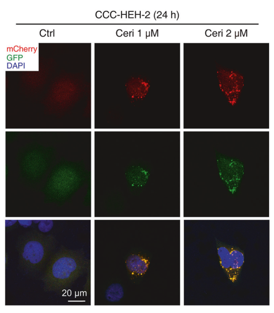
Confocal images of the Ad-mCherry-GFP-LC3 reporter system in CCC-HEH-2 cells
Research Background
Cardiovascular toxicity associated with cancer therapy remains a major clinical challenge. Ceritinib is an anaplastic lymphoma kinase (ALK) inhibitor associated with cardiovascular adverse events, but its mechanisms are not fully understood. Recently, Zhifei Xu's team at the Innovation Institute for Artificial Intelligence in Medicine of Zhejiang University published a study entitled AKT (Ser473) suppression mediates ceritinib cardiotoxicity through autophagic flux impairment and mitochondrial injury in Acta Pharmacologica Sinica (IF 10.4), revealing that AKT suppression drives ceritinib cardiotoxicity through autophagic flux impairment and mitochondrial injury, and positioning AMPK-driven, TFEB-related lysosomal restoration as a mechanism-based cardioprotective strategy that is independent of AKT recovery.
Research Results
1. Ceritinib induces cardiomyocyte apoptosis in vivo and in vitro
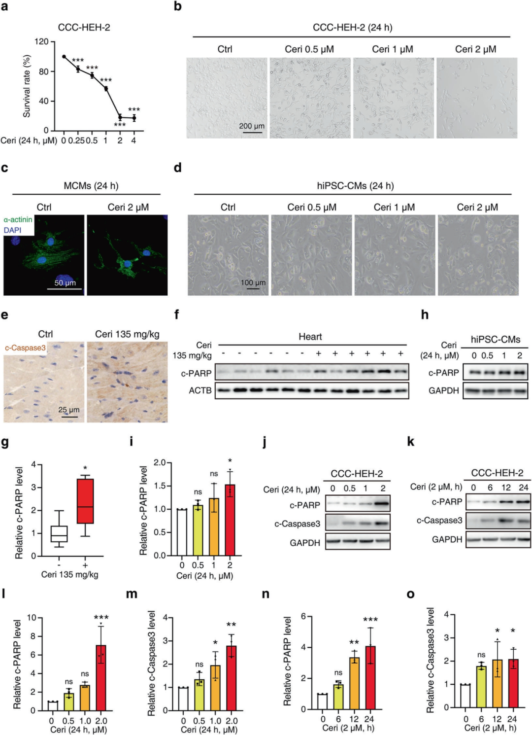
The authors found that ceritinib induced systolic dysfunction and myocardial injury in mice, and further explored whether apoptosis is involved in this process. Ceritinib treatment enhanced the immunohistochemical signal of cleaved caspase-3 (c-Caspase3) in the myocardium and markedly elevated cleaved poly(ADP-ribose) polymerase (c-PARP), indicating caspase-dependent apoptosis. In vitro, ceritinib reduced cell viability and worsened cell morphology, including cell shrinkage, detachment and reduced density, in CCC-HEH-2 cells. Western blotting confirmed dose- and time-dependent increases in c-PARP and c-Caspase3, and flow cytometry further confirmed dose- and time-dependent accumulation of apoptotic cells. In hiPSC-CMs, ceritinib dose-dependently increased c-PARP accompanied by morphological deterioration, and in primary mouse cardiomyocytes α-actin immunofluorescence revealed pronounced structural damage after ceritinib treatment. These results indicate that ceritinib activates caspase-dependent apoptosis in cardiomyocytes in a dose- and time-dependent manner across multiple experimental models.
Figure 1. Ceritinib induces cardiomyocyte apoptosis in vivo and in vitro
2. Ceritinib suppresses autophagy at the lysosomal degradation step
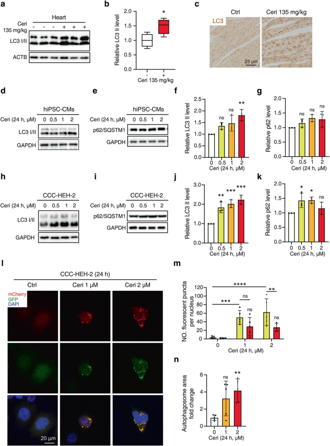
Western blotting showed marked accumulation of LC3-II in ceritinib-treated hearts and cardiomyocytes, whereas steady-state p62 levels did not increase in parallel with LC3-II, ruling out a definitive interpretation of autophagic flux by immunoblotting alone. The authors therefore used the Ad-mCherry-GFP-LC3 reporter system for direct assessment of autophagic flux and lysosomal function, and found that ceritinib increased both the number and the area of yellow LC3-positive puncta, indicating accumulation of incompletely acidified LC3-positive compartments and impaired late autophagic processing. In addition, lysosomal acidification was impaired in primary mouse cardiomyocytes and CCC-HEH-2 cells, and in hiPSC-CMs pro-CTSD accumulation with a decreased mature/pro-CTSD ratio supported lysosomal acidification defects and impaired cathepsin D maturation. In ceritinib-treated hiPSC-CMs, LAMP1 protein levels increased, and LAMP1 immunofluorescence showed a corresponding increase in CCC-HEH-2 cells, indicating compensatory lysosomal membrane expansion in response to the functional impairment. These results indicate that ceritinib impairs autophagic flux mainly at the lysosomal degradation step rather than at autophagosome-lysosome fusion. Mechanistic studies revealed that the AKT-GSK3β signalling branch contributes to the lysosomal enzyme maturation defects and mitochondrial protein loss caused by ceritinib exposure.
Figure 2. Ceritinib induces LC3-II accumulation and late-stage autophagy impairment in cardiomyocytes
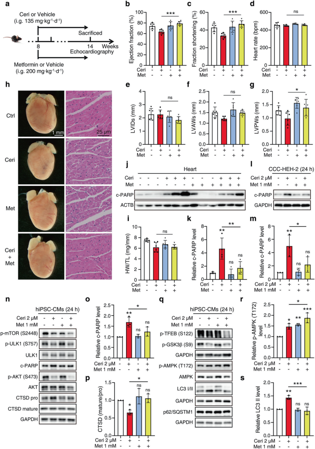
Given that metformin activates AMPK, which extensively interacts with the AKT signalling pathway, the authors explored whether metformin could ameliorate ceritinib-induced myocardial injury. In a mouse model receiving metformin and ceritinib in combination, combination therapy significantly improved cardiac function, reduced cardiac c-PARP levels and ameliorated myocardial structural damage. In vitro, metformin co-treatment significantly reduced ceritinib-induced c-PARP accumulation. Mechanistically, ceritinib treatment reduced p-mTOR (Ser2448); metformin also lowered p-mTOR, and the combination produced stronger inhibition than either agent alone. Metformin co-treatment elevated p-AMPK (Thr172) and reduced p-TFEB (Ser122), consistent with activation of an AMPK-driven, TFEB-related lysosomal repair programme. Metformin also reduced pro-CTSD accumulation, restored the mature/pro-CTSD ratio and decreased LC3-II accumulation, indicating improved lysosomal proteolytic capacity and relief of late autophagic stress. Importantly, metformin did not reverse the ceritinib-induced suppression of p-AKT (Ser473) or p-GSK3β (Ser9), demonstrating that its protective effect is independent of AKT-GSK3β restoration. These data indicate that metformin improves lysosomal function through an AMPK-driven mechanism, thereby alleviating ceritinib-induced cardiotoxicity independently of AKT-GSK3β reactivation.
This study demonstrates that ceritinib induces cardiotoxicity by suppressing AKT (Ser473) phosphorylation, creating a mismatch between enhanced autophagy initiation and defective lysosomal degradation that ultimately leads to autophagic flux impairment and mitochondrial injury. Metformin alleviates this toxicity through an AMPK-driven, TFEB-related mechanism of lysosomal function restoration. Overall, these findings define the AKT-autophagy-lysosome-mitochondria circuitry underlying ceritinib cardiotoxicity and provide a rationale for a mechanism-based cardioprotective strategy for cancer patients receiving ALK inhibitors.