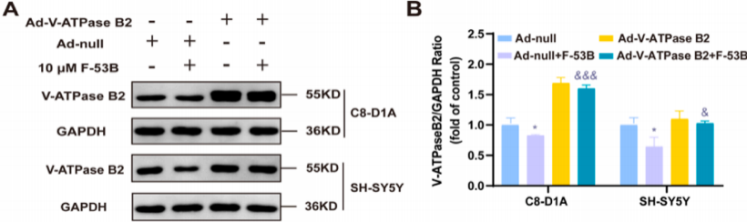
V-ATPase B2 expression increased significantly in C8-D1A and SH-SY5Y cells after Ad-V-ATPase B2 intervention
Research Background
6:2 chlorinated polyfluoroalkyl ether sulfonate (F-53B), a substitute for perfluorooctane sulfonate (PFOS), is widely distributed in various natural environments. F-53B is considered neurotoxic, but its mechanism of action remains unclear. On 4 January 2025, the research team of Qiang Niu and Mulatibieke Keerman at the School of Medicine, Shihezi University, published in Journal of Hazardous Materials (IF 12.2) a paper entitled F-53B disrupts energy metabolism by inhibiting the V-ATPase-AMPK axis in neuronal cells, which elucidated a new mechanism by which F-53B induces neurotoxicity through the V-ATPase-AMPK axis and indicated that V-ATPase and AMPK are potential therapeutic targets.
Research Results
1. F-53B inhibits V-ATPase B2 expression and V-ATPase activity and impairs mitochondrial function in neuronal cells
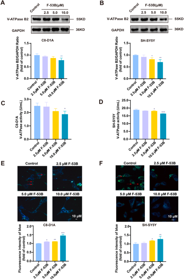
Mitochondrial ROS content and mitochondrial membrane potential (MMP) were measured by immunofluorescence, and it was found that, compared with the control group, F-53B exposure impaired mitochondrial function and reduced ATP levels in neuronal cells. Compared with the control group, F-53B exposure inhibited p-AMPK expression in C8-D1A and SH-SY5Y cells, and the protein levels of PFKFB3 and NDUFS1 also decreased significantly with increasing F-53B dose. Complex I (CI) activity was further analysed in the two cell lines, and exposure to 10.0 μM F-53B significantly inhibited CI activity. Western blot analysis showed that F-53B significantly inhibited V-ATPase B2 expression and reduced V-ATPase activity in C8-D1A and SH-SY5Y cells. In addition, lysosomal pH was elevated in F-53B-treated cells. Further analysis showed that F-53B exposure significantly inhibited TRPML1 and CaMKK2 protein expression in C8-D1A and SH-SY5Y cells in a dose-dependent manner, and increased Ca2+ accumulation in lysosomes.
F-53B exposure increases lysosomal pH and inhibits V-ATPase B2 expression and V-ATPase activity in neuronal cells
2. V-ATPase B2 overexpression alleviates F-53B-induced energy metabolism impairment
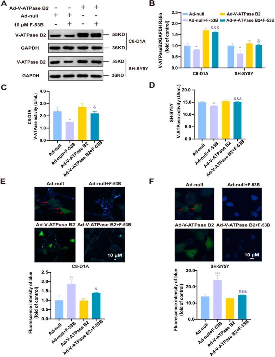
The research team used an adenoviral vector to overexpress V-ATPase B2 in C8-D1A and SH-SY5Y cells to verify whether upregulation of V-ATPase B2 could counteract the above F-53B-induced damage. The results showed that in the Ad-V-ATPase B2 + F-53B combination group, V-ATPase B2 expression increased and V-ATPase activity was significantly elevated; overexpression of V-ATPase B2 effectively restored the F-53B-induced increase in lysosomal pH. In addition, in the combination group, TRPML1 expression increased markedly, lysosomal Ca2+ accumulation decreased, and CaMKK2 and p-AMPK protein expression as well as ATP levels were all significantly elevated, indicating that V-ATPase B2 overexpression alleviated the F-53B-induced impairment of energy metabolism. Further studies demonstrated that respiratory syncytial virus (RSV) alleviated F-53B-induced suppression of p-AMPK expression and mitochondrial damage, restoring the energy metabolism pathway in neuronal cells.
V-ATPase B2 overexpression restored the F-53B-induced decrease in V-ATPase B1 expression and V-ATPase activity and alleviated the increase in lysosomal pH in neuronal cells
Conclusions
The results of this study show that F-53B inhibits V-ATPase activity, elevates lysosomal pH, leads to lysosomal Ca2+ accumulation, and subsequently blocks p-AMPK and its downstream signalling pathways by reducing CaMKK2 expression. This regulatory mechanism disrupts the V-ATPase-AMPK signalling axis, causing severe energy metabolism dysfunction in neuronal cells and reducing ATP production. Notably, intervention with Ad-V-ATPase B2 and RSV significantly attenuated F-53B-induced neurocytotoxicity. These findings provide new insights into the mechanism of F-53B neurotoxicity and offer potential targets for future neuroprotective strategies aimed at modulating the V-ATPase-AMPK signalling axis.