Acta Pharmacologica Sinica | Qizhu Tang team at Renmin Hospital of Wuhan University reveal that SRPK3 regulates mTOR/P70 S6K-mediated lethal autophagy through KLF3 Ser71 phosphorylation and aggravates myocardial ischemia-reperfusion injury
Adenovirus and lentiviral vectors for SRPK3 myocardial ischemia-reperfusion study | Qizhu Tang team at Renmin Hospital of Wuhan University, Acta Pharmacologica Sinica (IF 10.4)
Paper title: SRPK3 exacerbates cardiac ischemia/reperfusion injury by regulating KLF3-mTOR/P70 S6K-mediated autophagy
Journal: Acta Pharmacologica Sinica (IF 10.4)
Collaborating partner: Renmin Hospital of Wuhan University - Professor Qizhu Tang's team
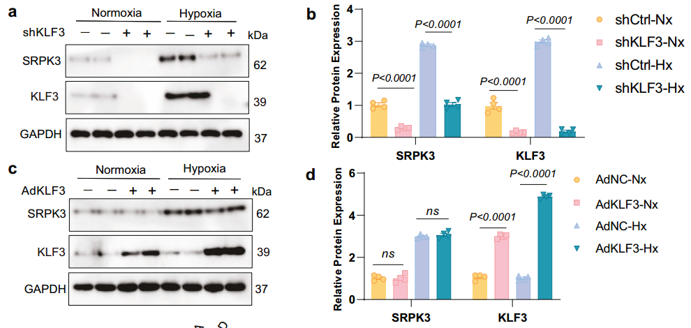
WB showing successful overexpression or knockdown of KLF3
Research Background
Myocardial ischemia-reperfusion (I/R) injury remains one of the leading causes of cardiac dysfunction and death worldwide. Serine/arginine-rich protein kinase 3 (SRPK3) is highly expressed in the myocardium, but its specific pathological role in I/R injury has not been fully elucidated. Recently, Professor Qizhu Tang's team at Renmin Hospital of Wuhan University published in Acta Pharmacologica Sinica (IF 10.4) a paper entitled SRPK3 exacerbates cardiac ischemia/reperfusion injury by regulating KLF3-mTOR/P70 S6K-mediated autophagy, systematically elucidating that SRPK3 induces lethal autophagy through the KLF3-mTOR axis and thus acts as a detrimental regulator in myocardial I/R injury. These findings establish the SRPK3-KLF3 interaction as a key molecular switch and provide a promising therapeutic target for ischaemic heart disease.
Research Results
1. SRPK3 promotes ischaemia-hypoxia-induced acute responses through the mTOR/P70 S6K signalling pathway
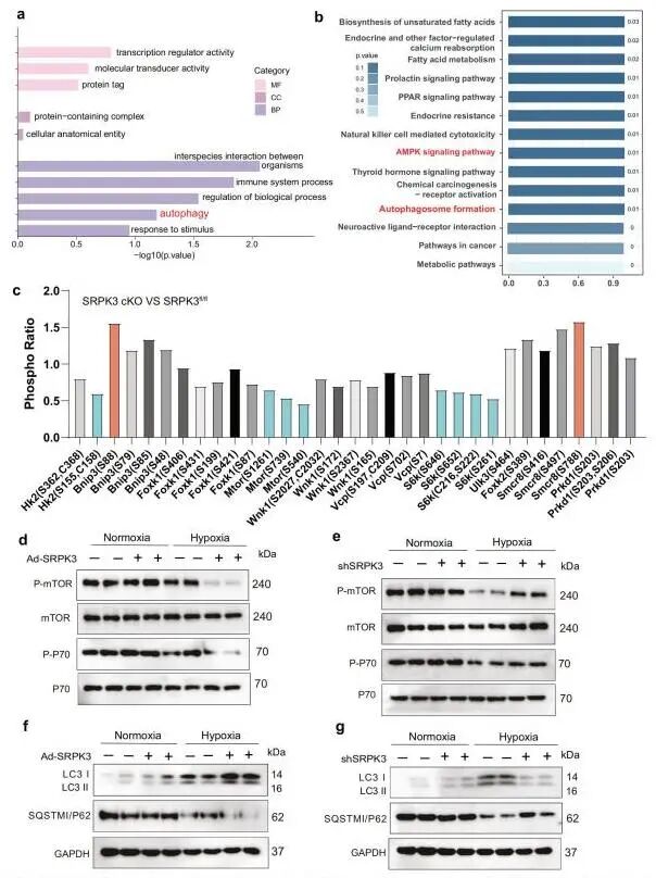
The results showed that SRPK3 was significantly upregulated in myocardial I/R injury and was mainly expressed in cardiomyocytes. Overexpression of SRPK3 in cardiomyocytes aggravated myocardial ischaemia, oxidative stress and apoptosis in the early stage of ischemia-reperfusion injury, and further worsened cardiac dysfunction and myocardial remodelling in the late stage of injury; cardiomyocyte-specific knockout of SRPK3 alleviated both early and late myocardial injury after ischemia-reperfusion, mainly by inhibiting apoptosis, reducing oxidative stress and improving pathological adverse remodelling. The research team further explored the downstream signalling pathways through which SRPK3 regulates the acute-phase response to ischaemia-hypoxia. Combining 4D label-free proteomics and phosphoproteomics, GO and KEGG enrichment indicated that autophagy is the core biological process of SRPK3. Time-gradient detection in the early stage of ischemia-reperfusion revealed that changes in SRPK3 protein levels were synchronised with changes in the autophagy markers LC3-II and p62, suggesting that SRPK3 regulates autophagic flux in the acute phase. Results from a mTOR signalling phosphorylation array showed that SRPK3 knockout promoted phosphorylation of key mTOR activation sites and other sites of ribosomal protein S6 kinase (S6K) in the early stage of ischemia-reperfusion injury. Cell function experiments further confirmed that SRPK3 mediates excessive autophagy under ischaemia-hypoxia stimulation by inhibiting the mTOR/P70 S6K signalling pathway, driving the acute-phase injury response of the myocardium.
SRPK3 promotes ischaemia-hypoxia-induced acute responses through the mTOR/P70 S6K signalling pathway
2. Effect of SRPK3-KLF3 Ser71 on the mTOR/P70 S6K signalling pathway
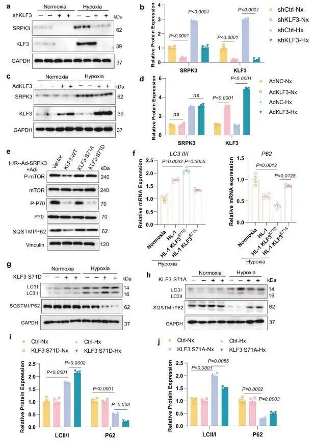
Studies of the molecular mechanism showed that SRPK3 can bind to the transcription factor KLF3 and phosphorylate KLF3 at Ser71; phosphorylation at this site enhances the SRPK3-KLF3 interaction, promotes nuclear localisation of p-KLF3 Ser71, and thereby regulates myocardial injury-related signalling pathways. Given that KLF3, as a transcriptional repressor, can mediate selective autophagic degradation, the research team explored the interaction between SRPK3 and KLF3 in the downstream mTOR signalling cascade. Knockdown of KLF3 using adenovirus downregulated SRPK3 protein levels, whereas overexpression of KLF3 did not change SRPK3 expression, indicating a unidirectional regulatory relationship between KLF3 and SRPK3. RT-qPCR results showed that SRPK3 overexpression significantly reduced mTOR mRNA levels, and KLF3 knockdown significantly reversed this inhibition; the phosphorylation-deficient mutant KLF3 S71A failed to inhibit mTOR mRNA, indicating that this transcriptional repression depends on SRPK3-mediated phosphorylation at Ser71. Using different KLF3 mutants in hypoxia/reoxygenation cell rescue experiments, the authors further confirmed that SRPK3 phosphorylates KLF3 at Ser71, inhibits mTOR transcription, downregulates mTOR/P70 S6K signalling pathway activity, and ultimately aggravates excessive autophagy in cardiomyocytes under hypoxic conditions.
SRPK3-KLF3 Ser71 affects the mTOR/P70 S6K signalling pathway
Conclusions
This study systematically revealed the dual role of SRPK3 in ischemia-reperfusion injury: it is both a driver of myocardial injury and a key regulator of autophagic imbalance. SRPK3 mediates KLF3 Ser71 phosphorylation and regulates the mTOR signalling axis, inhibits protective autophagy, aggravates apoptosis and oxidative stress, and ultimately causes deterioration of cardiac function. This study not only elucidates the pathological mechanism of ischemia-reperfusion injury but also lays a theoretical foundation for developing precise therapeutic strategies targeting SRPK3.