

Research Background
Renal fibrosis caused by chronic kidney disease is the main cause of end-stage renal failure (uraemia). Studies have shown that the TGF-β1 (transforming growth factor-β1) signalling pathway plays an important role in the process of renal interstitial fibrosis. Under TGF-β stimulation, phosphorylation of Smad3 (a signal transducer protein) is a key step in the activation of the entire TGF-β/Smad3 pathway. Previous research focused mainly on genes encoding key proteins — that is, mutations in conventional key genes alter the proteins they encode and thereby cause abnormal biological function. However, with the development of research into molecular biological mechanisms, it has become apparent that epigenetics also plays an important role in the development of renal interstitial fibrosis.
Research Approach
The researchers used TGF-β for induction and, through high-throughput screening, examined the expression of differentially expressed long non-coding RNAs (lncRNAs) in renal tubular epithelial cells, finding that lnc-TSI expression increased significantly under TGF-β stimulation. Functionally, the researchers found that lnc-TSI negatively regulates the TGF-β/Smad3 signalling pathway by specifically inhibiting Smad3 phosphorylation. Mechanistic studies revealed that lnc-TSI can bind the MH2 domain of Smad3 and thereby prevent the binding of TGF-β type I receptor to Smad3.
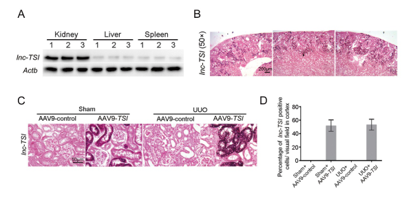
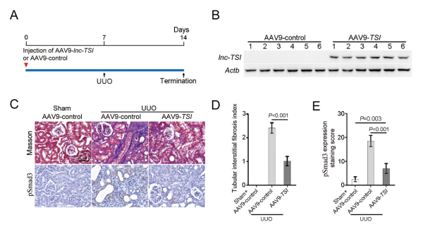
Animal experiments showed that overexpressing the human gene lnc-TSI in the mouse kidney via adeno-associated virus AAV9 effectively prevented the onset and progression of renal fibrosis in a mouse model. In a cohort study of patients with chronic kidney disease, the expression level of lnc-TSI in kidney biopsy tissue was negatively correlated with the severity of renal interstitial fibrosis. In patients with chronic kidney disease who were followed up long-term and underwent repeat kidney biopsies, the researchers found that patients with lower renal lnc-TSI expression at the first biopsy had more severe renal fibrosis and decline in renal function 4 years later than those with higher lnc-TSI expression.
Viral Product and Administration Method
- Viral product
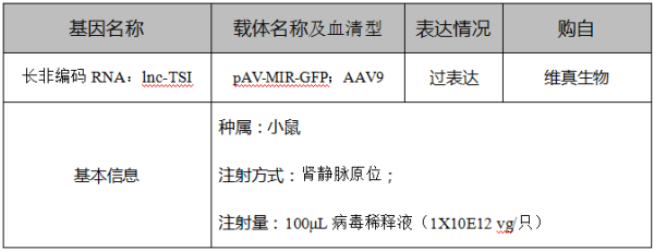
- AAV9-TSI (WZBIO)
- Target tissue / organ
- Mouse kidney
- Model
- Unilateral ureteral obstruction (UUO)
- Injection route
- In situ injection into the renal vein
- Viral titre
- 1×10E14 vg/mL
- Dose per mouse
- 100 μL (10 μL of AAV9-TSI diluted to 100 μL with saline)
- Timing
- Injected one week before modelling; mice sacrificed one week after modelling
Procedure
The mouse renal vein was clamped with an arterial clip, a 31G needle was used to inject into the renal vein, and the clip was kept closed for 15 minutes before being released.
Analysis of Experimental Results