How much do you know about autophagy?
1.Introduction to Cellular Autophagy
Cellular autophagy (autophagy = self-eating) refers to the process by which eukaryotic cells, under the regulation of autophagy-related genes (Atg), utilize lysosomes to degrade their own cytoplasmic proteins and damaged organelles. Cellular autophagy prevents cellular damage, promotes cell survival under conditions of nutrient deprivation, and responds to toxic stimuli. It includes basal autophagy under physiological conditions and induced autophagy under stress conditions. The former is a self-protective mechanism that benefits cell growth and development, protects cells from metabolic stress and oxidative damage, and plays an important role in maintaining intracellular homeostasis as well as in the synthesis, degradation, and recycling of cellular products. However, excessive autophagy can lead to metabolic stress, degradation of cellular components, and even cell death. Studies have shown that cellular autophagy can play an important role in various physiological and pathological processes such as cell homeostasis, aging, immunity, tumorigenesis, and neurodegenerative diseases.
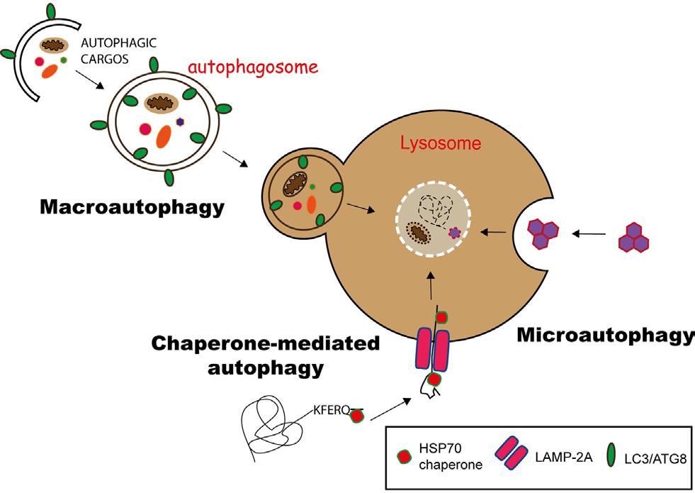
Cellular autophagy can be divided into three types based on the different materials being enclosed and the mode of transport (Figure 1):
① Macroautophagy: This involves the formation of an autophagosome with a double membrane structure to enclose intracellular material, which is then fused with a lysosome. The term cellular autophagy generally refers to macroautophagy.
② Microautophagy: Specific organelles are engulfed directly through deformations on the surface of lysosomes or vacuoles.
③ Chaperone-mediated autophagy (CMA): Proteins with KEFRQ-like motifs are transported to lysosomes via the LAMP-2A transporter with the assistance of HSP70 chaperones.
Figure 1. Three autophagy pathways
(Duraes Fernanda V et al. Front Immunol, 2015)
2.The process of autophagy in cells
Autophagy is an evolutionarily conserved intracellular catabolic process in which large molecules in the cytoplasm, aggregated proteins, damaged organelles, or pathogens are transported to lysosomes and degraded by lysosomal hydrolases, producing nucleotides, amino acids, fatty acids, sugars, and adenosine triphosphate, which are ultimately recycled back into the cytoplasm. Various factors such as starvation, radiation, hypoxia, bacterial invasion, and growth factor deprivation can induce the occurrence of cell autophagy.
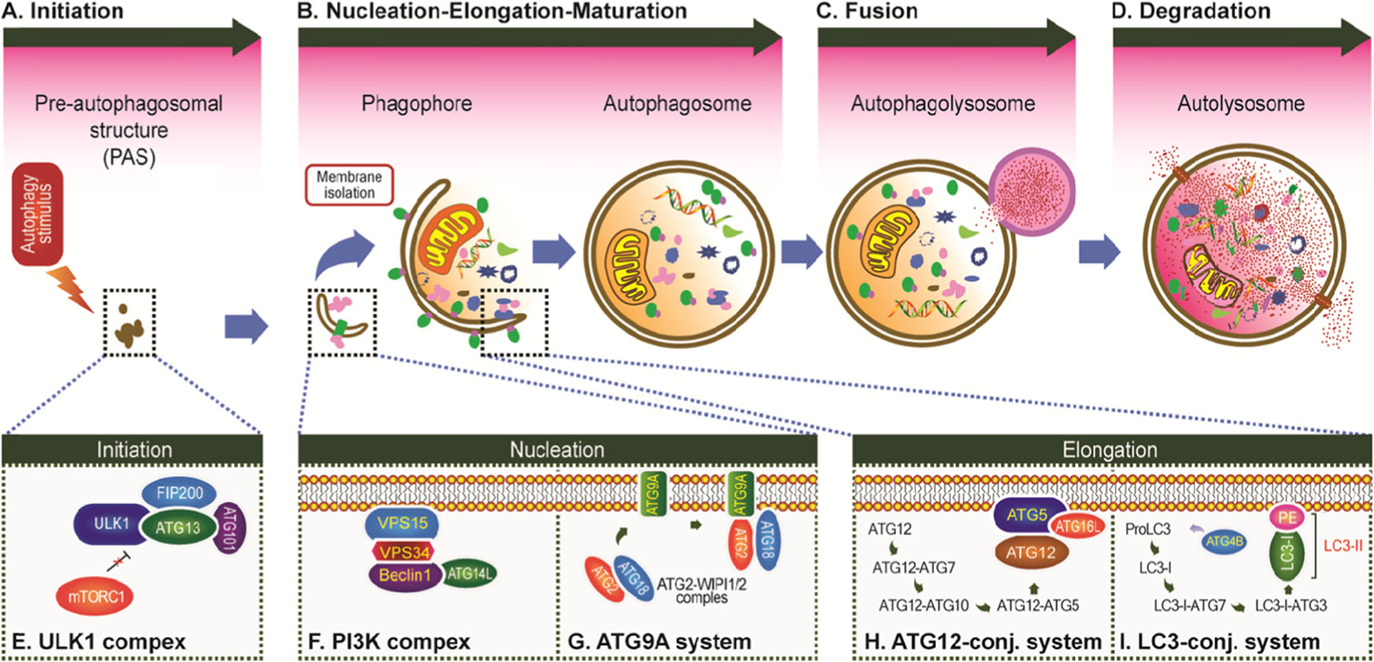
The process of cellular autophagy can be broadly divided into the following four stages (Figure 2):
①Initiation of Cellular Autophagy Under the regulation of cellular autophagy induction signals, the ULK1 complex and various ATG proteins are activated and localized at the pre-autophagosomal structure.
②Formation of Isolation Membrane and Autophagosome ATG proteins and lipids continue to be recruited, forming a cup-shaped double-membrane structure (isolation membrane, phagophore); as the isolation membrane gradually extends, it completely encloses the cytoplasmic components to be degraded, eventually forming a closed autophagosome.
③Fusion of Autophagosome with Lysosome After the formation of the autophagosome, its cargo is transported to the lysosome through the intracellular transport system and fused with it.
④Lysis of Autophagosome The fusion of the autophagosome with the lysosome forms an autolysosome, which ultimately degrades its cargo under the action of lysosomal hydrolases.
In summary, the essence of cellular autophagy is actually membrane rearrangement within cells. During this process, a closed vesicle with a double membrane that encapsulates random or specific substrates is formed, namely the autophagosome, which then fuses with the lysosome to form an autolysosome and degrade the substrate.
Figure 2. The process of autophagy in cells (Li et al., Molecular Cancer, 2020)
3.Cell Autophagy-Related Proteins
The occurrence of cell autophagy is related to nutritional status, energy state, oxidative stress, ischemia and hypoxia, etc., and it is regulated by multiple mechanisms such as ULK1 pathway, Beclin1 pathway, AMPK pathway, etc. During the process of cell autophagy occurrence, there are various cell autophagy-related proteins that can regulate and control different stages of cell autophagy formation.
As shown in Figure 2, during the process of cell autophagy occurrence, various cell autophagy-related genes regulate different stages of the cell autophagy flux. To date, scientists have identified more than 40 genes encoding ATG proteins in yeast, and most of them are highly conserved between yeast and mammals. In mammalian cells, starvation-induced cell autophagy is regulated by about 20 core ATG genes, which are continuously recruited near the vacuole and assembled to form the precursors of cell autophagosomes. The classification and functions of these genes (Table 1) are as follows:
①ULK1 Kinase Core Complex: Includes ULK1/2, ATG13, RB1CC1/FIP200, and ATG101;
②PI3K Complex: Includes VPS34, VPS15, Beclin1, and ATG14L;
③ATG9A Transport System: Includes ATG9A, WIPI1/2, and ATG2A;
④ATG12 Ubiquitin-like Binding System: Includes ATG12, ATG7, ATG10, ATG5, and ATG16L1;
⑤LC3 Ubiquitin-like Binding System: Includes LC3A/B/C, ATG7, ATG3, and ATG4A/B/C/D.
4.Regulation of Cellular Autophagy
Cellular autophagic activity at the basal level is very low, making it unsuitable for observation. Therefore, research on cellular autophagy often requires artificial intervention.
The mTOR (mechanistic target of rapamycin) kinase plays an important regulatory role in the cellular autophagy response. When activated by mTOR kinase, the cell autophagy response is inhibited; while when mTOR (AMPK and p53 signaling) is inhibited, the mechanism of cellular autophagy is initiated. Autophagy-related genes (Atg) regulate the formation of cellular autophagosomes by forming Atg12-Atg5 and LC3-II (Atg8-II) complexes.
Autophagy can both inhibit and promote apoptosis, and the two reactions are widely present in organisms. Under conditions of nutrient deficiency, autophagy acts as a pro-survival mechanism for cells, but excessive autophagy can also lead to cell death. However, cell death caused by autophagy is morphologically distinct from apoptosis. Several pro-apoptotic signaling factors, such as TNF, TRAIL, and FADD, can also induce autophagic responses. Additionally, Bcl-2 is an important regulator of autophagy; it is an anti-apoptotic protein that inhibits Beclin-1-induced autophagy by forming a complex with Beclin-1.
A detailed list of some of the reported intervention drugs is provided in Table 1:
|
Cell Autophagy Inducer
|
autophagy antagonist
|
|
Rapamycin: Rapamycin, mTOR inhibitor (most commonly used)
|
Chloroquine: Chloroquine, lysosome inhibitor
|
|
EBSS: Earle's Balanced Salt Solution, induces starvation
|
Bafilomycin A1: Bafilomycin A1, proton pump inhibitor
|
|
Lithium Chloride: Lithium chloride, IMPase inhibitor
|
3-Methyladenine: 3-Methyladenine, hVps34 inhibitor
|
|
Xestospongin B/C: Xestospongin B/C, IP3R blocker etc
|
NH4Cl: Ammonium chloride, lysosome inhibitor etc
|
Table 1. Cell Autophagy Agonists and Antagonists
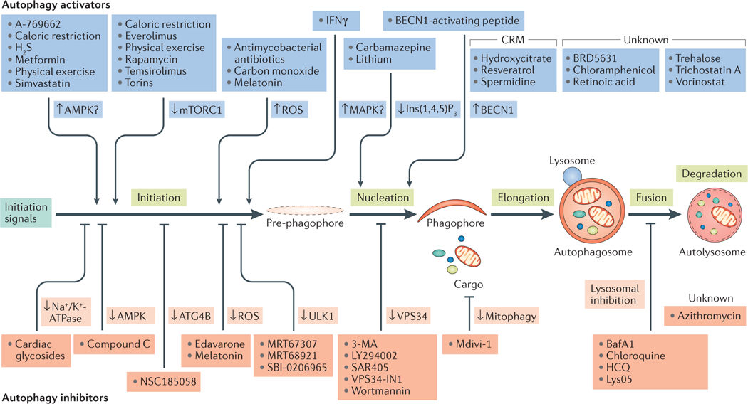
Commonly used cell autophagy activators include Rapamycin, EBSS, etc., and commonly used cell autophagy inhibitors include Chloroquine, 3-MA, NH4Cl, and Bafilomycin A1, etc. These cell autophagy activators and inhibitors can activate/inhibit different stages of cell autophagy occurrence respectively, so researchers can select appropriate activators or inhibitors according to the needs of the experiment. For more drugs that regulate the cell autophagy pathway, see Figure 3.
Figure 3. Drug regulation of the autophagic pathway
(Lorenzo Galluzzi et al. Nat Rev Drug Discov, 2017)
5、Detection Methods of Cellular Autophagy
Under physiological conditions, the activity of cellular autophagy is usually low. However, upon stimuli such as starvation, hypoxia, and diseases, the activity of cellular autophagy will be significantly upregulated. In addition, the inhibition of cellular autophagy is also associated with certain diseases, such as cancer, neurodegenerative diseases, and infectious diseases. Given the close relationship between cellular autophagy and various physiological and pathophysiological processes, it is necessary for scientists to select an ideal detection method for cellular autophagy. After cells are induced or inhibited for autophagy, the commonly used observation and detection methods are:
5.1 Transmission Electron Microscopy Method
Autophagosomes belong to subcellular structures and cannot be seen under ordinary light microscopy. Directly observing the morphological changes of autophagy at different stages under transmission electron microscopy is a very direct method. The morphological characteristics of each stage of autophagy are shown in Table 2; The morphology of autophagosomes and autolysosomes in cells under transmission electron microscopy is shown in Figure 4.
|
Autophagic stage
|
Morphological characteristics
|
|
Isolation membrane
|
Crescent-shaped or cup-shaped, with a double or multilayered membrane, tends to surround cytoplasmic components.
|
|
Autophagosome
|
A liquid-filled, vesicular structure with a double or multilayer membrane, containing cytoplasmic components such as mitochondria, endoplasmic reticulum, ribosomes, etc.
|
|
Autophagic lysosome
|
A monolayer with degraded cytoplasmic components.
|
Table 2. Characteristics of the various stages of cellular autophagy
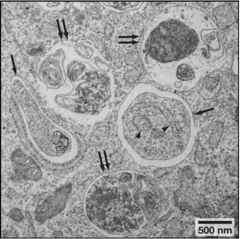
Figure 4. Morphology of autophagic vacuoles (single arrow) and autolysosomes (double arrow) observed under transmission electron microscopy.
(Noboru, Mizushima et al. Cell, 2010)
5.2 Observation Method with Fluorescence Microscope
LC3 (light chain 3), also known as MAP1LC3 (microtubule-associated protein light chain 3), is involved throughout the entire cellular autophagy process and is currently recognized as a marker for cell autophagy. In mammals, LC3 shares homology with the yeast autophagy-related protein Apg8/Aut7/Atg8. There are three types of LC3 in mammals: LC3A, LC3B, and LC3C. Among these, LC3B is the most widely used. After synthesis, the carboxyl terminus of LC3 is cleaved by Atg4 to remove the last five amino acids, exposing a glycine residue, resulting in cytosolic-localized LC3-I. During the process of cellular autophagy, LC3-I undergoes modification and processing by ubiquitin-like systems, including Atg7 and Atg3, and conjugates with phosphatidylethanolamine (PE) to form LC3-II, which is localized on the inner and outer membranes of the cell autophagosome. After fusion of the cell autophagosome with the lysosome, LC3-II on the outer membrane is cleaved by Atg4, generating LC3-I for recycling; LC3-II on the inner membrane is degraded by lysosomal enzymes, resulting in low levels of LC3 within the cell autophagolysosomes (Figure 5). Therefore, researchers can detect cell autophagy occurrence by observing endogenous LC3 or GFP-LC3 using fluorescence microscopy.
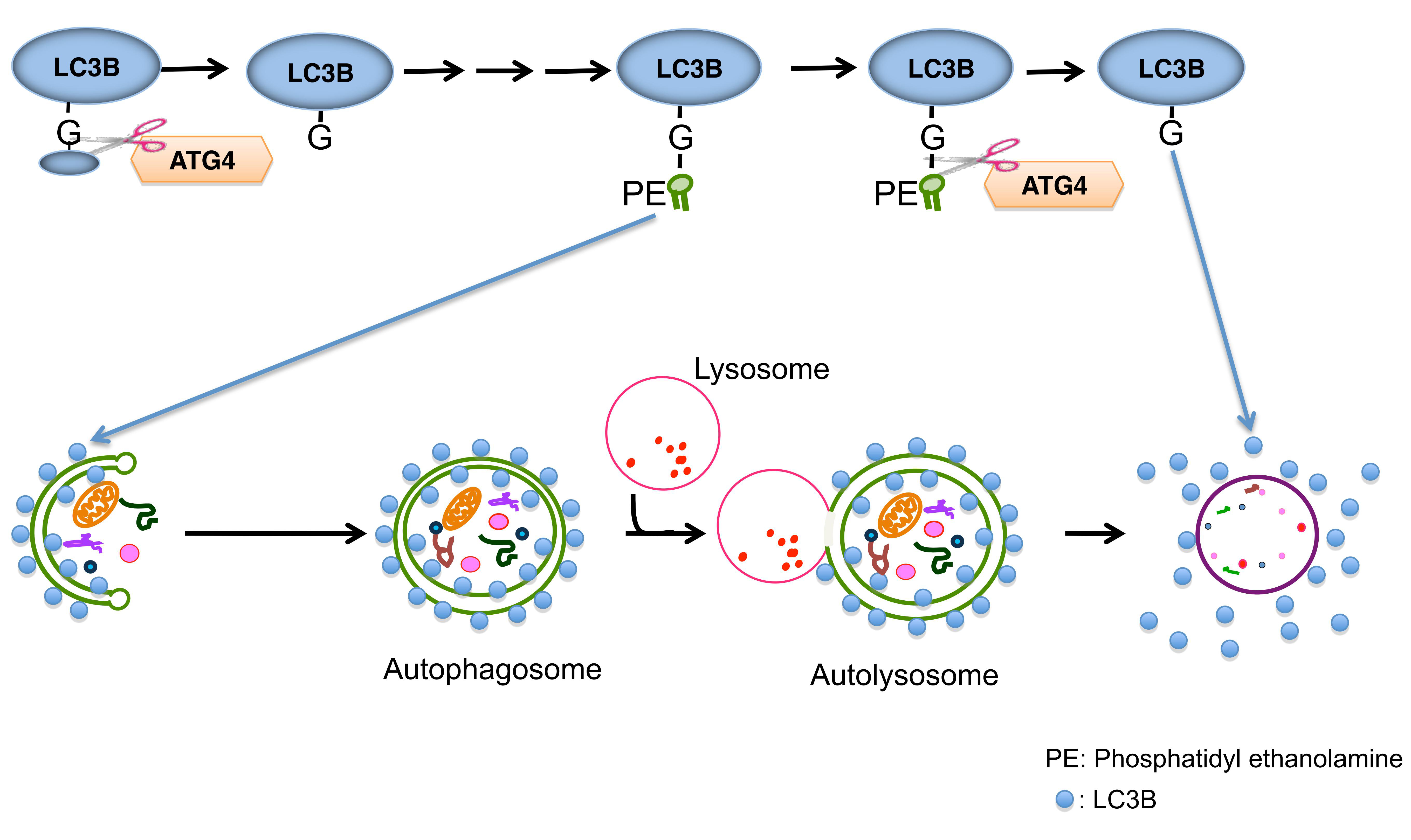
Figure 5. Pathway of LC3 in Cell Autophagy Occurrence
(Kiriyama, Y et al. International Electronic Conference on Medicinal Chemistry, 2016)
The GFP-LC3 single fluorescent and mCherry-GFP-LC3 double fluorescent labeling systems
During the formation of autophagosomes, the GFP-LC3 or mCherry-GFP-LC3 fusion proteins are transferred to the membranes of autophagic vesicles, forming multiple bright green or yellow fluorescent spots under fluorescence microscopy. After the formation of autolysosomes, the acidic lysosomal environment quenches the GFP fluorescence, while the mCherry fluorescence remains unaffected, resulting in red fluorescence from the autolysosomes (Figure 6). Therefore, researchers can monitor the autophagic flux using the LC3 fluorescent labeling system.
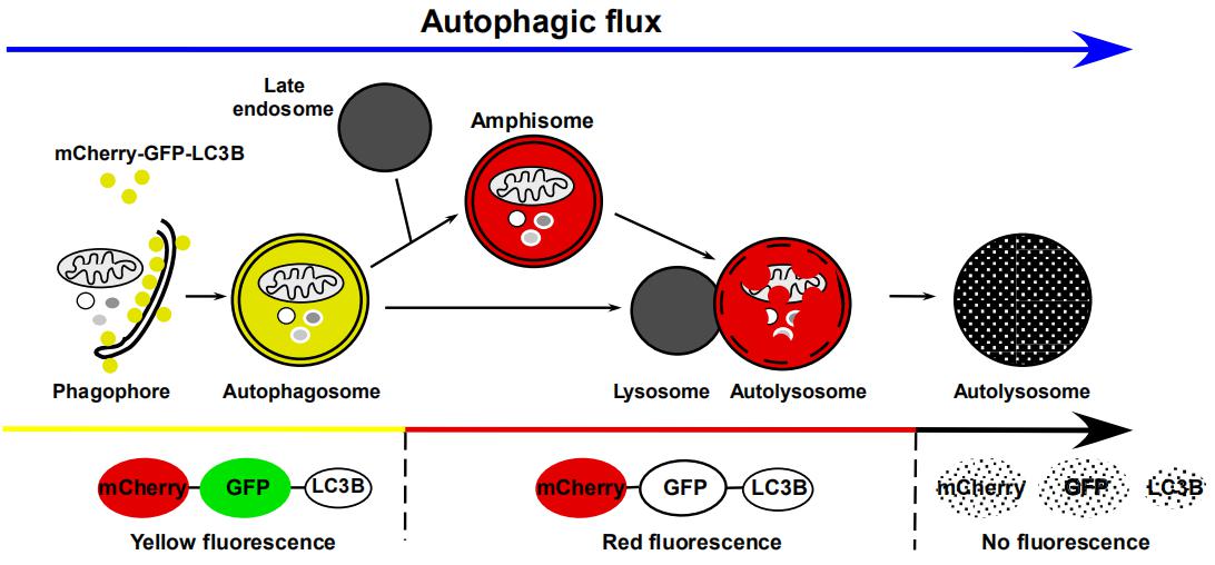
Figure 6. Tracking different stages of the autophagic flux in LC3-expressing cells using a dual-labeling system
(Hansen TE, Johansen TB. BMC Biology, 2011)
5.3 Western Blot detection of LC3 and p62 protein expression levels
① Use Western Blot to detect changes in the LC3-II/I ratio to evaluate cell autophagy formation (Figure 7). When cell autophagy forms, cytoplasmic LC3-I is enzymatically cleaved by a small peptide segment and then combines with PE to transform into membrane-type LC3-II. Therefore, the size of the LC3-II/I ratio can be used to estimate the level of cell autophagy.
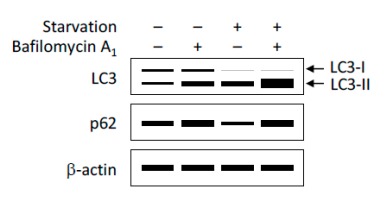
Figure 7. Schematic representation of the results from Western blot analysis for LC3 and p62 protein expression.
(Yoshii, S.R. and N. Mizushima. Int J Mol Sci, 2017.)
In addition to LC3, changes in the expression levels of other cellular autophagy substrates can also be used to monitor the flux of cellular autophagy. Among them, p62 is a widely studied cellular autophagy substrate. p62 (also known as SQSTM1 protein) consists of three structural domains: the N-terminal Phox and Bem1 (PB1) domain, the zinc finger domain, and the C-terminal ubiquitin associated (UBA) domain. Studies have shown that the linker region (LRS region) between the zinc finger domain and UBA domain of the p62 protein is responsible for binding with the cellular autophagy receptor protein Atg8/LC3, while the UBA domain is responsible for recruiting ubiquitinated proteins. During the formation of cellular autophagosomes, p62 acts as a bridge linking LC3 and polyubiquitinated proteins, selectively encapsulated into cellular autophagosomes, and then degraded by proteases in cellular autophagolysosomes (Fig. 8), so the expression level of p62 protein is negatively correlated with cellular autophagy activity. Therefore, detecting the expression level of p62 protein by Western Blot can also be used to evaluate the level of cellular autophagy (Fig. 7).
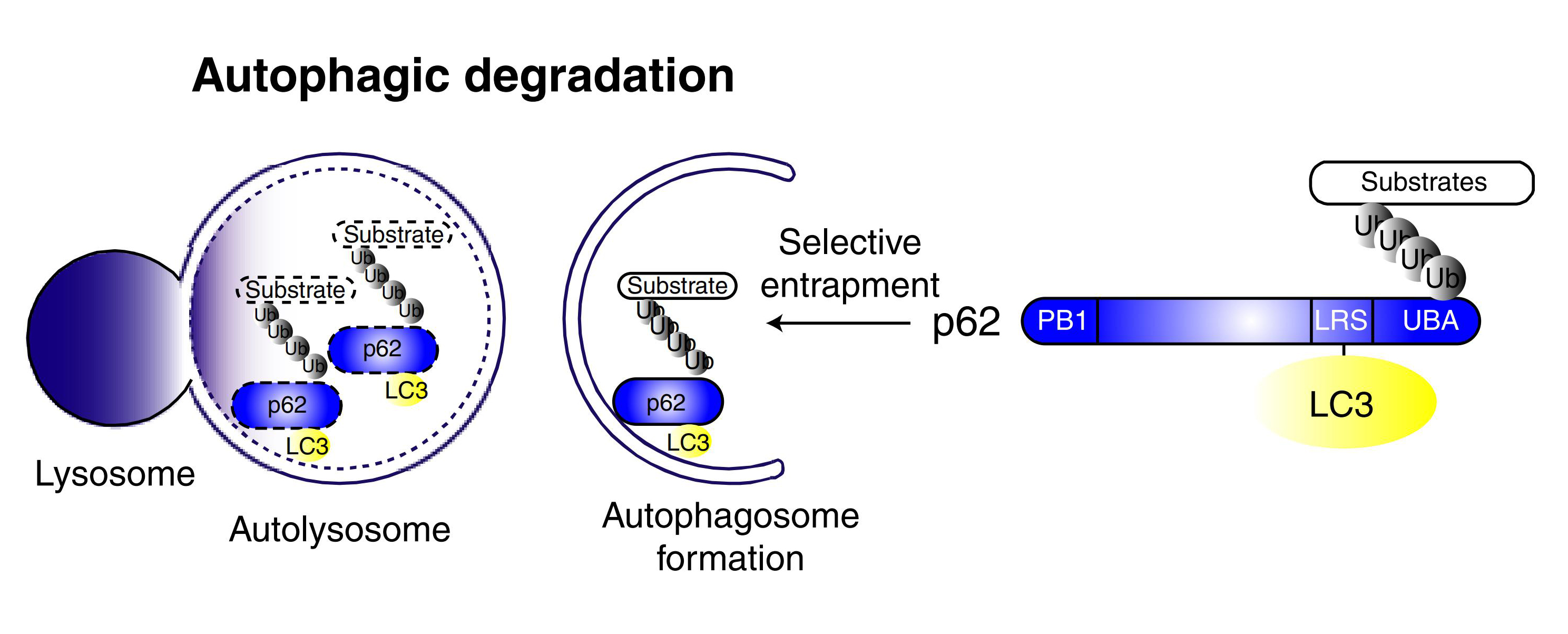
Figure 8. A selective autophagy model mediated by p62
(Ichimura Y et al. J Biol Chem, 2008)
5.4 Evaluation of Cellular Autophagy Based on Keima Protein
Keima is a pH-sensitive fluorescent protein whose dual-peak excitation spectrum depends on the surrounding pH value, activating at 440 nm and 586 nm in neutral and acidic environments, respectively. The different fluorescence signals of Keima under neutral and acidic pH can intuitively reflect the degree of autophagy in cells. Transferring cytoplasmic Keima to lysosomes can reflect non-selective cell autophagy, while fusing Keima with specific proteins (such as fusion with mitochondrial targeting sequence - mt-Keima, serving as a marker for mitochondrial cell autophagy) can be used to reflect selective cell autophagy. It's worth noting that detection based on Keima cannot be performed on fixed cells, as this detection entirely relies on the acidity of lysosomes.
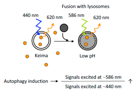
Figure 9. Evaluation of cellular autophagy based on Keima protein
(Yoshii, S.R. and N. Mizushima. Int J Mol Sci, 2017.)
6. Weizhen Cell Autophagy-Related Products
Weizhen provides tools for cell autophagy research, including a large number of ready-to-use gene clones related to cell autophagy, shRNA clones, CRISPR clones, as well as LC3 cell autophagy single and double label clones and viruses. Multiple strategies, various specifications, and some ready-to-use products are available to meet your diverse research needs.
6.1 Viral Products
|
Product Name
|
Fluorescent protein
|
Titer
|
|
AdV5-CMV-GFP-LC3
|
GFP
|
≥1*10E10pfu/ml
|
|
AdV5-CMV-mCherry-GFP-LC3
|
mCherry、GFP
|
≥1*10E10pfu/ml
|
|
AAV-CMV-GFP-LC3
|
GFP
|
≥1*10E13vg/ml
|
|
AAV-CMV-mCherry-GFP-LC3
|
mCherry、GFP
|
≥1*10E13vg/ml
|
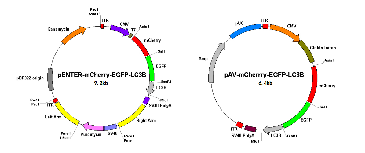
Figure 10. Schematic diagram of the Vero cell autophagy double-labeling virus vector
6.2 Cloned Products
Partial Cell Autophagy-Related Genes
|
Product Code
|
Gene name
|
Gene Number
|
|
CH852478
|
alfy
|
NM_014991
|
|
CH808275
|
ulk1
|
NM_003565
|
|
CH858197
|
sqstm1
|
NM_003900
|
|
CH808956
|
sqstm1
|
NM_001142298
|
|
CH815035
|
p150
|
NM_005407
|
|
CH801228
|
fip200
|
NM_014781
|
|
CH895682
|
p150
|
NM_012414
|
|
CH885062
|
ampk
|
NM_017431
|
|
CH842000
|
ampk
|
NM_002733
|
|
CH818994
|
ampk
|
NM_006253
|
|
CH801722
|
ampk
|
NM_006252
|
|
CH805185
|
ampk
|
NM_206907
|
|
CH888389
|
pi3k
|
NM_001142633
|
|
CH841329
|
p150
|
NM_014602
|
|
CH823236
|
pi3k
|
NM_002649
|
|
CH864199
|
pi3k
|
NM_005026
|
|
CH814123
|
pi3k
|
NM_006219
|
|
CH800089
|
pi3k
|
NM_006218
|
|
CH892216
|
pi3k
|
NM_004570
|
|
CH816369
|
pi3k
|
NM_002646
|
|
CH824737
|
nbr1
|
NM_031862
|
|
CH800308
|
mtor
|
NM_004958
|
|
CH872072
|
lamp2
|
NM_001122606
|
|
CH846728
|
lamp2
|
NM_013995
|
|
CH876641
|
lamp1
|
NM_005561
|
|
CH806988
|
p150
|
NM_001206557
|
|
CH807619
|
rubicon
|
NM_001145642
|
|
CH832873
|
p150
|
NM_014633
|
|
CH860614
|
p150
|
NM_005483
|
|
CH859985
|
bcl-2
|
NM_000633
|
|
CH871029
|
atg7
|
NM_006395
|
|
CH871778
|
atg7
|
NM_001136031
|
|
CH879325
|
atg7
|
NM_001144912
|
|
CH828790
|
atg5
|
NM_004849
|
|
CH800065
|
atg4
|
NM_032885
|
|
CH804672
|
atg4
|
NM_178221
|
|
CH896350
|
atg4
|
NM_013325
|
|
CH883248
|
atg4
|
NM_178326
|
|
CH878793
|
atg4
|
NM_052936
|
|
CH853753
|
atg4
|
NM_178270
|
|
CH811236
|
atg3
|
NM_022488
|
|
CH882182
|
atg16l1
|
NM_017974
|
|
CH802102
|
atg16l1
|
NM_198890
|
|
CH807003
|
atg16l1
|
NM_001190266
|
|
CH878363
|
atg16l1
|
NM_001190267
|
|
CH886537
|
atg16l1
|
NM_030803
|
|
CH838864
|
atg14
|
NM_014924
|
|
CH842100
|
atg13
|
NM_014741
|
|
CH833706
|
atg13
|
NM_001205122
|
|
CH871057
|
atg13
|
NM_001205120
|
|
CH897141
|
atg13
|
NM_001205119
|
|
CH821707
|
atg12
|
NM_004707
|
|
CH805558
|
atg10
|
NM_031482
|
|
CH805050
|
p150
|
NM_001150
|
|
CH882903
|
pras40
|
NM_032375
|
|
CH879027
|
p150
|
NM_007313
|
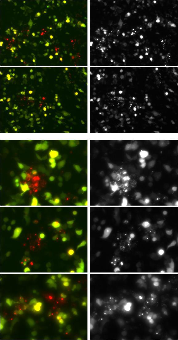
Attachment: Effect of infection with the Weizhen Cell Autophagy Dual-Label System on inducing cell autophagy in HEK293 cells.