The activity of neurons is the foundation of complex behavioral manifestations in the brain. And the activity of neurons can cause rapid changes in intracellular free calcium concentration. Calcium ions, as an important intracellular signaling molecule, play a crucial role in neuronal function, such as the release of neurotransmitters in synaptic vesicles. Calcium imaging technology can directly measure the dynamic calcium flow in neurons and neuronal tissues, thereby achieving the goal of detecting neuronal activity.
1. Calcium imaging principle
Using special fluorescent dyes or calcium ion indicators, the changes in calcium ion concentration in neurons are expressed through fluorescence intensity. Through rapid imaging of calcium ion changes, the activity changes of neurons are reflected.
2、Calcium Indicators
Calcium indicators are essential tools for studying the function of calcium ions. According to the differences in fluorescence spectra, calcium ion affinity, and chemical properties, they can be roughly divided into two categories: chemical indicators and genetic coding indicators.
Chemical indicators are traditional tools for detecting the concentration of calcium ions in the cytoplasm, mainly small molecules that can specifically bind to calcium ions, including Fura-2, indo-1, fluo-3, fluo-4, Calcium Green-1, etc. These small molecules are calcium chelating agents called BAPTA, which is a homologue of ethylene glycol tetraacetic acid (EGTA) and has high selectivity for Ca2+. Chemical fluorescent indicators are usually delivered to the target site by electrode penetration or by means of acetoxymethyl ester and dextrose.
Genetically encoded calcium indicators (GECIs) are a new type of calcium indicator that has emerged with the development of genetic engineering technology. They can achieve long-term and real-time dynamic detection of calcium ions in vivo experiments, and can also characterize the changes in calcium ions in certain subcellular structures using specific localization signals from organelles.
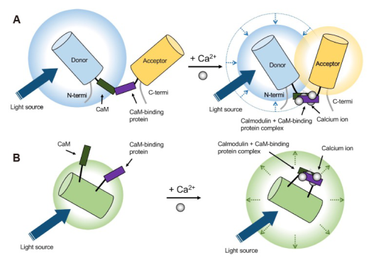
According to the principle of luminescence, GECIs can be divided into two categories: GECIs based on single fluorescent proteins and GECIs composed of fluorescent protein pairs through fluorescence resonance energy transfer (FRET). The former mainly includes Pericams, GCaMP, and Camgaroos, while the latter mainly includes TN-XXL, Camelerons, D3cpV, etc. GECIs calcium indicators are mainly introduced into the target site through viral infection.
Genetic encoded calcium ion indicator
(Jihae Oh et al. Korean J Physiol Pharmacol. 2019)
3、GCaMPs calcium indicator
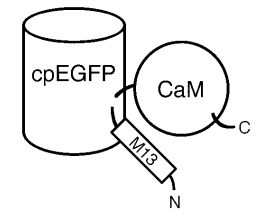
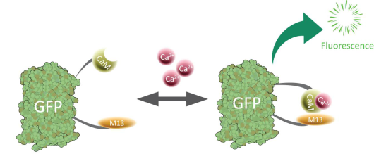
Among GECIs, GCaMP with structure oriented design is the most widely used. GCaMPs are products of the fusion of a single green fluorescent protein GFP, calmodulin CaM, and smooth muscle cell myosin light chain kinase fragment M13. In the presence of Ca2+, CaM binds to M13, greatly increasing the intensity of fluorescence. The schematic diagram of its structure and imaging principle are shown in the following figure.
|
|
|
|
Schematic diagram of GCaMPs
|
structure Principle of GCaMPs imaging
|
4、Calcium imaging research steps
Taking genetic coding calcium ion indicators as an example, briefly describe the research steps:
Step 1: Select appropriate GECIs calcium ion indicators;
Step 2: Introduce the calcium ion indicator into the target location through viral infection;
Step 3: Record the dynamic changes of calcium ions and analyze neuronal activity.
5、How to choose a suitable calcium indicator (taking GCaMPs as an example)
Since the emergence of GCaMP1 in 2001, scientists have continuously optimized its affinity, signal-to-noise ratio, dynamics, and dynamic range through structure based mutation and neuron based screening methods. They have successfully developed GCaMP2, GCaMP3, GCaMP5, GCaMP6, and jGCaMP7 to meet the different imaging experiments of researchers. So, how should we choose among the numerous GCaMPs calcium indicators? As is well known, different imaging experiments require the use of GCaMP with different properties. For example, GCaMP with low background fluorescence is suitable for wide field imaging technology and can simultaneously detect the activity of a large number of neurons. This is mainly due to the fact that a large number of neurons are labeled with GCaMP, which has a high number of photons and is prone to form stronger background fluorescence; High background fluorescence is suitable for detecting signals in neural subcellular structures, such as dendritic spines and axons, mainly due to their small volume, low GCaMP expression, low photon count, low background fluorescence, and even negligible effects; The fast dynamic GCaMP is suitable for detecting dynamic changes in neuronal firing patterns; The slow kinetics GCaMP is suitable for detecting all active neurons. The following table shows the characteristics of the GCaMP6 and jGCaMP7 series probes:
|
GCaMPs name
|
describe
|
|
GCaMP6s(slow)
|
Slow kinetics, suitable for detecting slow and sustained calcium signals
|
|
GCaMP6m(medium)
|
Moderate dynamics centered, suitable for detecting calcium signals with medium speed changes
|
|
GCaMP6f(fast)
|
Fast kinetics, suitable for detecting rapidly changing calcium signals
|
|
jGCaMP7s(sensitive)
|
For one action potential, Δ F/F0 increased by 5 times compared to 6s; slow dynamics
|
|
jGCaMP7f(fast)
|
For one action potential, Δ F/F0 increased by 2.5 times compared to 6s; Fast Dynamics
|
|
jGCaMP7b(bright)
|
For one action potential, Δ F/F0 increased threefold compared to 6s; High background fluorescence
|
|
jGCaMP7c(contrast)
|
Low background fluorescence
|
6、Reference:
1. Imaging and analysis of genetically encoded calcium indicators linking neural circuits and behaviors;
2. Ultrasensitive fluorescent proteins for imaging neuronal activity;
3. High-performance calcium sensors for imaging activity in neuronal populations and microcompartments.